Golfer’s Elbow (Medial Epicondylitis) and Steroid Injection Treatment: A Case Study
Medial epicondylitis, commonly known as golfer’s elbow, is a chronic condition characterized by pain and inflammation of the tendons that attach to the medial epicondyle of the elbow. This condition often results from repetitive wrist and forearm movements and can lead to significant discomfort and functional impairment. In this article, we present a case study of a 33-year-old woman diagnosed with golfer’s elbow.
Case presentation
Patient background:
- Age/gender: 33-year-old woman
- Symptoms: Right medial elbow pain persisting for one year following a minor trauma
- Previous treatment: Physical therapy and NSAIDs, with no significant improvement
The patient reported ongoing pain in the medial aspect of her right elbow, which significantly affected her daily activities. Despite conservative management, including physical therapy and NSAIDs, her symptoms persisted.
Diagnosis
Physical examination
- Observation: No redness or edema noted.
- Palpation: Tenderness over the medial epicondyle.
- Polk’s test: Positive for medial epicondylitis.
Laboratory parameters and imaging
- Fasting blood sugar: 100 mg%
- Erythrocyte sedimentation rate: 10 mm/hr
- Nerve conduction study: Normal
Ultrasound findings:
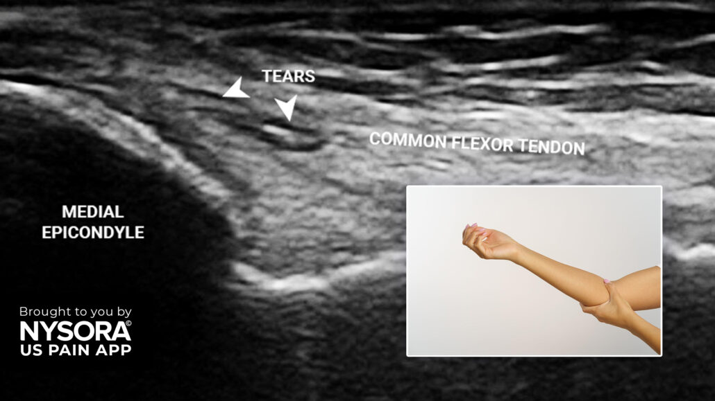
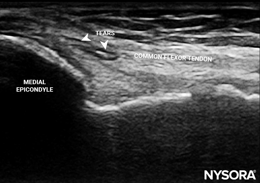
- Tendon tears: Minimal tears in the common flexor tendon at the insertion point onto the medial epicondyle.

Long axis view of the common flexor tendon and medial epicondyle. Note the tears at the insertion point.
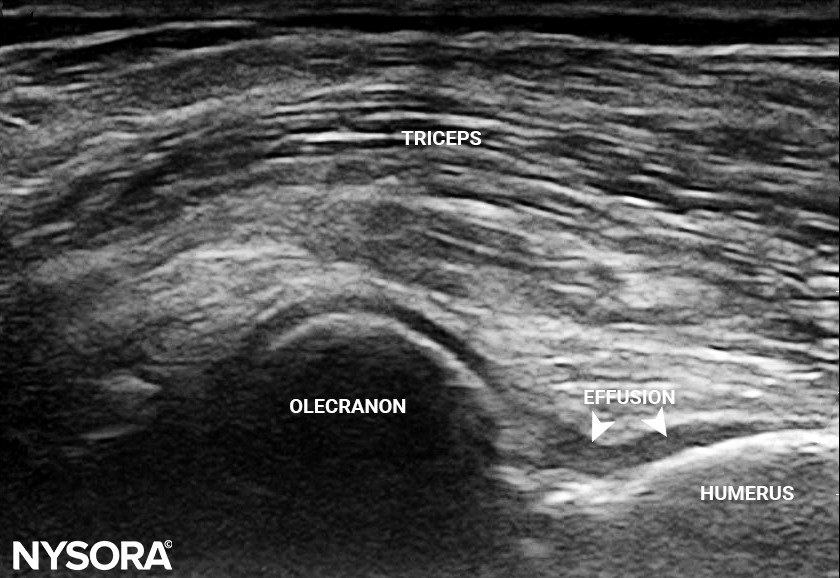
- Joint effusion: Small effusion in the anterior joint recess of the elbow.

Long axis view of the posterior elbow revealing a small effusion in the anterior joint recess.
Imaging summary:
- Diagnosis: Medial epicondylitis.
- Differential diagnoses: Pronator overuse syndrome, cubital tunnel syndrome, arthritis, cervical radiculopathy.
Treatment: Steroid injection
Steroid injection overview:
- Objective: Pain relief by reducing inflammation at the affected site.
- Injection site: Superficial to the common flexor tendon at its insertion onto the medial epicondyle.
Alternative treatments:
- Options: Dry needling, dextrose prolotherapy, botox, and PRP injection.
- Patient choice: The patient opted for a steroid injection for quicker pain relief, although PRP injection is the preferred long-term treatment.
Injection technique
Ultrasound setup:
- Transducer type: Linear, 3-13 MHz
- Preset: Musculoskeletal
- Orientation: Sagittal
- Depth: 2-3 cm
Patient position:
- Position: Supine with the shoulder abducted 90 degrees and the elbow flexed 90 degrees.
Needle insertion:
- Needle type: 26 gauge, 1.5-inch needle
- Technique: Inserted in-plane, superficial to the common flexor tendon to avoid tendon rupture.
Pharmacological agents:
- Lidocaine 2%
- Triamcinolone
Important note:
- Combination: Never combine steroids with PRP in the same treatment session.
Interested in more detailed information and the outcome for this patient? Download NYSORA’s US Pain App!
References
- Amin NH, Kumar NS, Schickendantz MS. Medial epicondylitis: evaluation and management. J Am Acad Orthop Surg. 2015;23(6):348-355.
- Tarpada SP, Morris MT, Lian J, Rashidi S. Current advances in the treatment of medial and lateral epicondylitis. J Orthop. 2018;15(1):107-110. Published 2018 Feb 2.


