Learning objectives
- Describe the etiology and causes of hypercapnia
- Diagnose hypercapnia
- Manage hypercapnia
Background
- Hypercapnia is defined as an elevation in the partial pressure of carbon dioxide (PaCO2) above 45 mmHg
- Due to the role of CO2 in Ph buffering, hypercapnia can lead to acid-base imbalances
Etiology
- Hypercapnia is secondary to disease rather than a single etiology
- Hypercapnia is caused by either increased metabolic CO2 production or respiratory failure
- Metabolic processes that increase CO2 production:
- Fever
- Thyrotoxicosis
- Increased catabolism in sepsis or steroid use
- Overfeeding
- Metabolic acidosis
- Exercise
- Respiratory failure: Failure to eliminate CO2 from the pulmonary system, hypoventilation secondary to decreased respiratory rate or decreased tidal volume
- Causes:
- Decreased central nervous system respiratory drive
- Anatomical defects
- Decreased neuromuscular response
- Increased dead space within the lung
- Causes:
- Hypercapnia can be acute or chronic, depending on the etiology
- Acute hypercapnia: PaCO2 >45 mmHg, HCO3 within normal range (~30 mmHg), resulting decrease in pH <7.35
- Chronic hypercapnia: Renal compensation, PaCO2 >45 mmHg, HCO3 elevated proportionally, less severe pH imbalance
Underlying pathologies
Pathologies that lead to hypercapnia include:
- Acute respiratory distress syndrome
- Asthma exacerbation
- Central or obstructive sleep apnea
- Chronic obstructive pulmonary disorder
- Drug overdose
- Exogenous CO2 inhalation
- Head or cervical cord injury
- Myasthenia gravis
- Myxedema
- Obesity-hypoventilation syndrome (Pickwickian syndrome)
- Polyneuropathy
- Poliomyelitis
- Primary muscle disorders
- Porphyria
- Primary alveolar hypoventilation
- Pulmonary edema
- Tetanus
Signs & symptoms
- Flushed skin
- Lethargy
- Inability to focus
- Mild headache
- Disorientation
- Dizziness
- Dyspnea
- Nausea
- Vomiting
- Fatigue
- Delirium
- Paranoia
- Depression
- Abnormal muscle twitches
- Palpitations
- Hyperventilation
- Hypoventilation
- Seizures
- Anxiety
- Syncope
Diagnosis
- Signs on physical examination may include:
- Fever
- Tachycardia
- Tachypnea
- Dyspnea
- Altered mental status
- Wheezing on auscultation
- Rales on auscultation
- Rhonchi on auscultation
- Decreased breath sounds
- hyper-resonant chest on percussion
- Increased anterior-posterior diameter of chest
- Cardiac murmur
- Signs of hypoxia
- Hepatosplenomegaly
- neurological deficit
- Confusion
- Somnolence
- Muscular weakness
- Peripheral edema
- Asterixis
- Papilledema
- superficial vein dilation
- Diagnostic testing:
- Complete blood count to determine anemia presence
- Complete metabolic panel (sodium, potassium, chloride, HCO3)
- Thyroid stimulating hormone to determine underlying hypo– or hyperthyroidism
- Arterial or venous blood gas (pH status, serum CO2, serum HCO3, anion gap)
- Spirometry (forced expiratory volume over 1 second, forced vital capacity)
- Chest X-ray
- Chest CT
- Echocardiography if cardiopulmonary abnormality is suspected
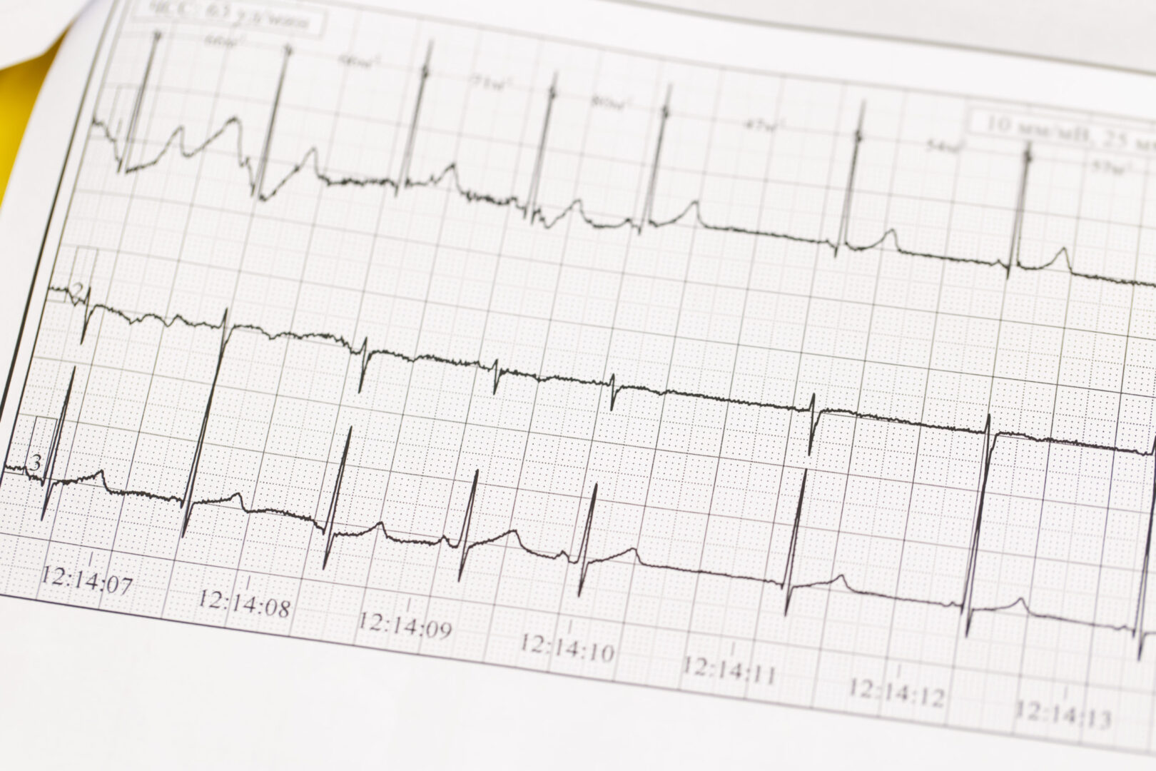
- ECG to evaluate central nervous system malfunctions
- Electromyography to evaluate neuromuscular disorders
- Polysomnography for suspected central or obstructive sleep apnea
Management
- Treat the underlying pathology
- Increase ventilation:
- Bi-level positive airway pressure
- Ventilation assist
- Continuous positive airway pressure ventilation
- Intubation with mechanical ventilation in critically ill patients
- Maintain oxygen saturation at 90% or higher
- Other options for critically ill ventilated patients:
- Increase minute ventilation
- Increase end-inspiratory pause prolongation
- Buffers: sodium bicarbonate, tromethamine
- Prone position ventilation
- Airway pressure release ventilation
- High-frequency oscillation ventilation
- Extracorporeal membrane oxygenation
- Low-flow extracorporeal CO2 removal devices
Suggested reading
- Rawat D, Modi P, Sharma S. Hypercapnea. [Updated 2022 Jul 25]. In: StatPearls [Internet]. Treasure Island (FL): StatPearls Publishing; 2022 Jan-. Available from: https://www.ncbi.nlm.nih.gov/books/NBK500012/
- Tiruvoipati R, Gupta S, Pilcher D, Bailey M. Management of hypercapnia in critically ill mechanically ventilated patients-A narrative review of literature. J Intensive Care Soc. 2020;21(4):327-333.
We would love to hear from you. If you should detect any errors, email us customerservice@nysora.com


