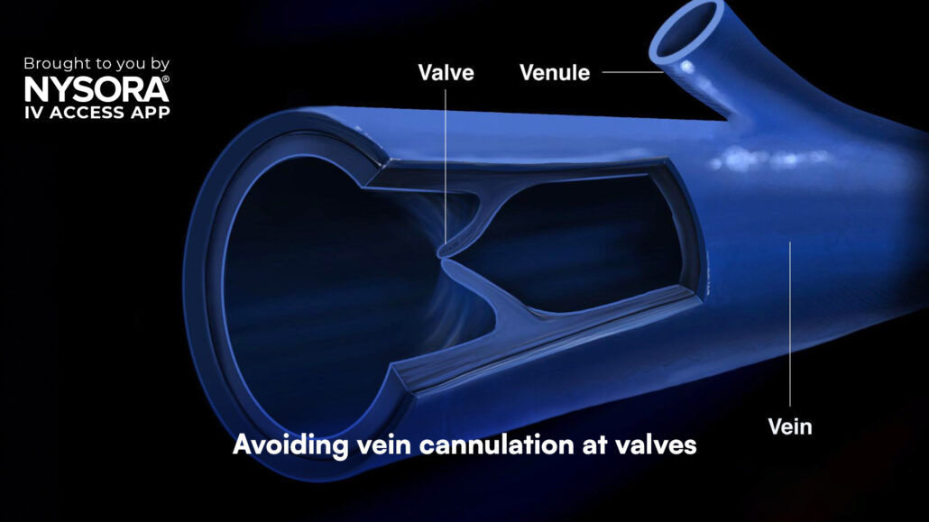
Avoiding vein cannulation at valves
When performing intravenous (IV) cannulation, one crucial consideration is the avoidance of venous valves. These small, flap-like structures within veins help regulate blood flow but can complicate IV access if not appropriately avoided.
Why avoid cannulation near venous valves?
Venous valves can present several challenges and risks during IV cannulation:
- Difficult insertion: Valves may obstruct the passage of the cannula, making insertion more challenging.
- Risk of damage: Puncturing or damaging a valve can lead to complications such as thrombophlebitis.
- Inefficient flow: A catheter tip positioned against a valve can impede the flow of fluids or medications.
- Increased risk of occlusion: Catheters near valves are more prone to becoming occluded.
- Patient discomfort: Cannulation at a valve site can be painful for patients.
- Potential for infiltration: Incorrect catheter placement due to a valve can lead to fluid leakage into surrounding tissues, causing swelling and discomfort.
- Reduced catheter longevity: Catheters placed near valves may need to be replaced more frequently, increasing patient discomfort and procedural frequency.
- Challenges in blood draw: Valves may obstruct the catheter and increase the risk of vein rupture if punctured.
Best practices for avoiding venous valves
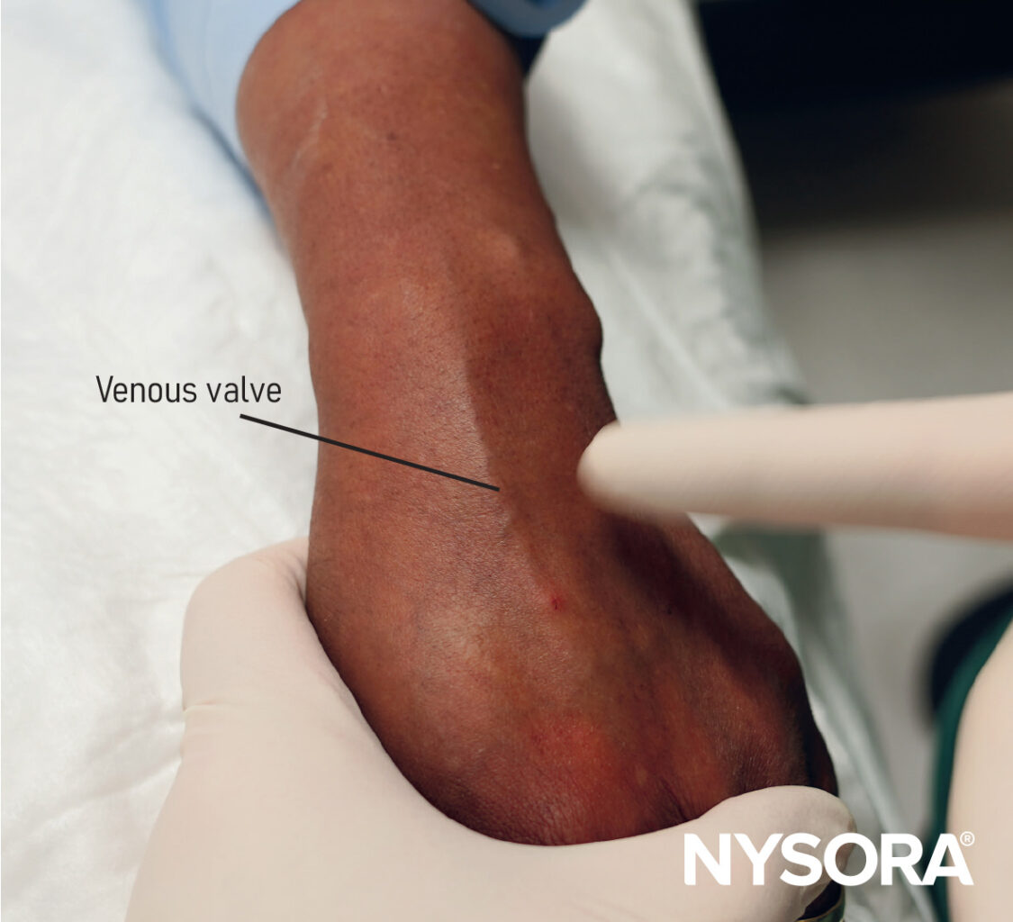
- Palpation
- Use your fingers to gently palpate the vein.
- Valves often feel like small “bumps” or “knots” along the vein.
- A noticeable bump followed by a depression may indicate the presence of a valve.
- Tourniquet Application
- Place a tourniquet above the site to distend the vein.
- This technique enhances visibility and palpability of veins, making valves easier to identify.
- Visual Inspection
- Look for visible signs of valves such as bifurcations or areas where the vein appears to branch or widen.
- Transillumination
- Use a vein light or transilluminator to make subcutaneous structures more visible.
- This tool highlights veins and reveals areas with potential valves.
- Ultrasound Guidance
- Use ultrasound to visualize the vein’s anatomy, including valves.
- Valves appear as bright (echogenic) structures that interrupt the vein’s lumen.
- This technique is particularly helpful in difficult IV placements.
- Assess Blood Flow
- Press distal to the vein and release. If the blood flow momentarily stops and resumes, it may indicate a valve.
- Training and Experience
- The ability to identify valves improves with experience and practice.
- Advanced training can enhance tactile and visual identification skills.
Conclusion
Avoiding venous valves during IV cannulation is a critical skill for healthcare professionals. By understanding their anatomy, recognizing their presence, and employing best practices, you can ensure safer and more effective IV access. Use tools such as palpation, tourniquet application, and ultrasound to identify valves, and rely on training and experience to refine your technique. Following these guidelines not only improves success rates but also enhances patient comfort and reduces complications.
Explore many resources, including easy-to-follow algorithms, expert tips, and clinical videos, on NYSORA’s IV Access App! It’s perfect for healthcare professionals at any level. Download today and start improving your IV catheterization techniques! All this valuable content is also available in NYSORA’s comprehensive IV manual on Amazon.


