Clinical case: Left ventricular function
Overview
Left ventricular function is pivotal in assessing the heart’s ability to pump blood effectively. Qualitative methods using point-of-care ultrasound (POCUS) allow clinicians to quickly differentiate between normal and reduced ventricular function, especially in undifferentiated shock states.
While quantitative assessments like ejection fraction are reserved for advanced echocardiography, POCUS provides an efficient qualitative approach to guide bedside management.
Goal
To assess left ventricular function and determine whether it is normal or reduced using key ultrasound views. This quick evaluation enables timely diagnosis and management of critical conditions.
Causes of left ventricular dysfunction
Left ventricular dysfunction may arise from several underlying pathologies, including:
- Coronary artery disease (CAD)
- Hypertension
- Left ventricular hypertrophy
- Valvular dysfunction (e.g., aortic regurgitation, mitral stenosis)
- Myocarditis
Key ultrasound views for assessing left ventricular function
To perform a qualitative assessment, the following 3 ultrasound views are commonly used:
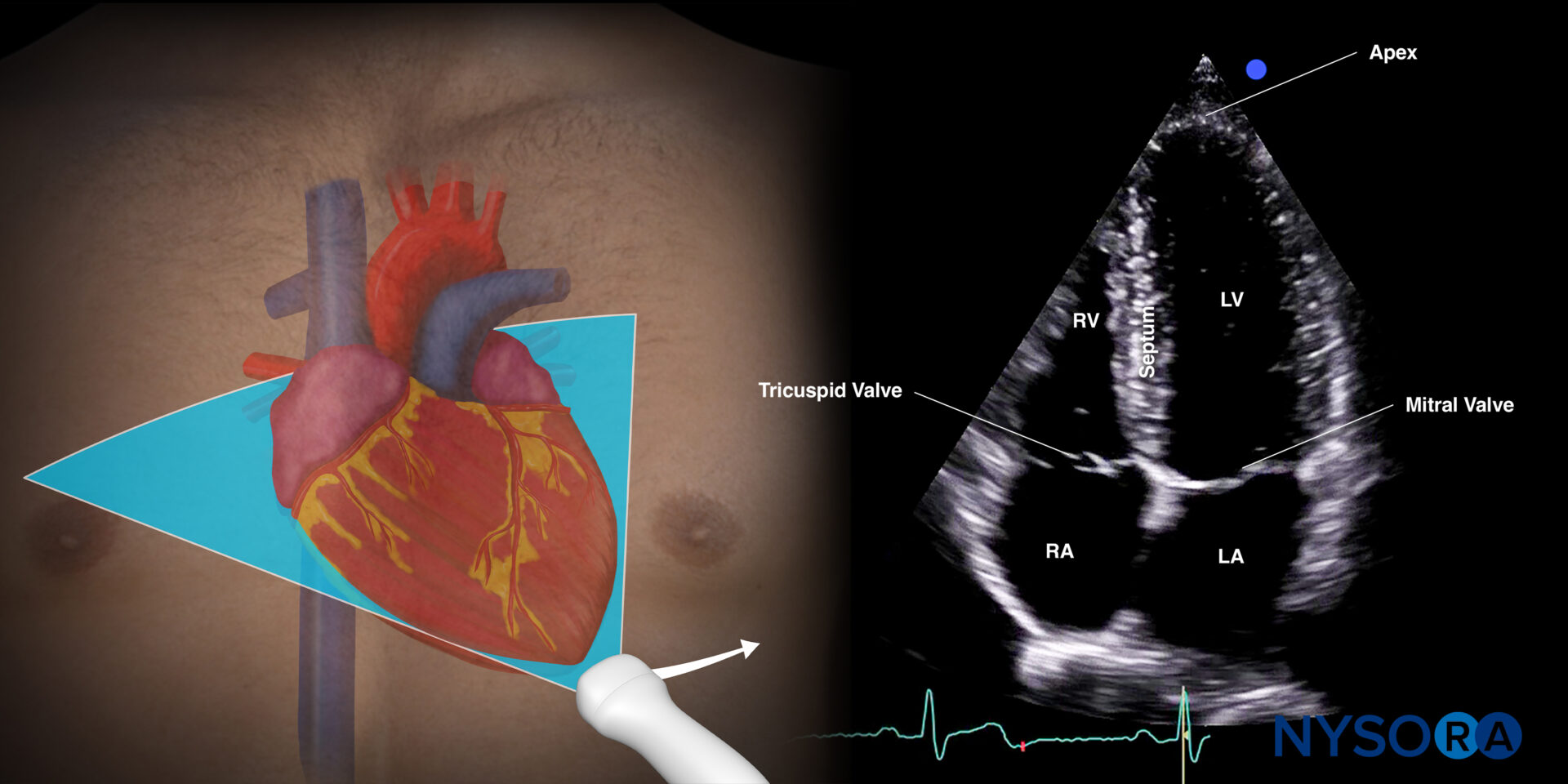
- Apical four-chamber (A4C) view
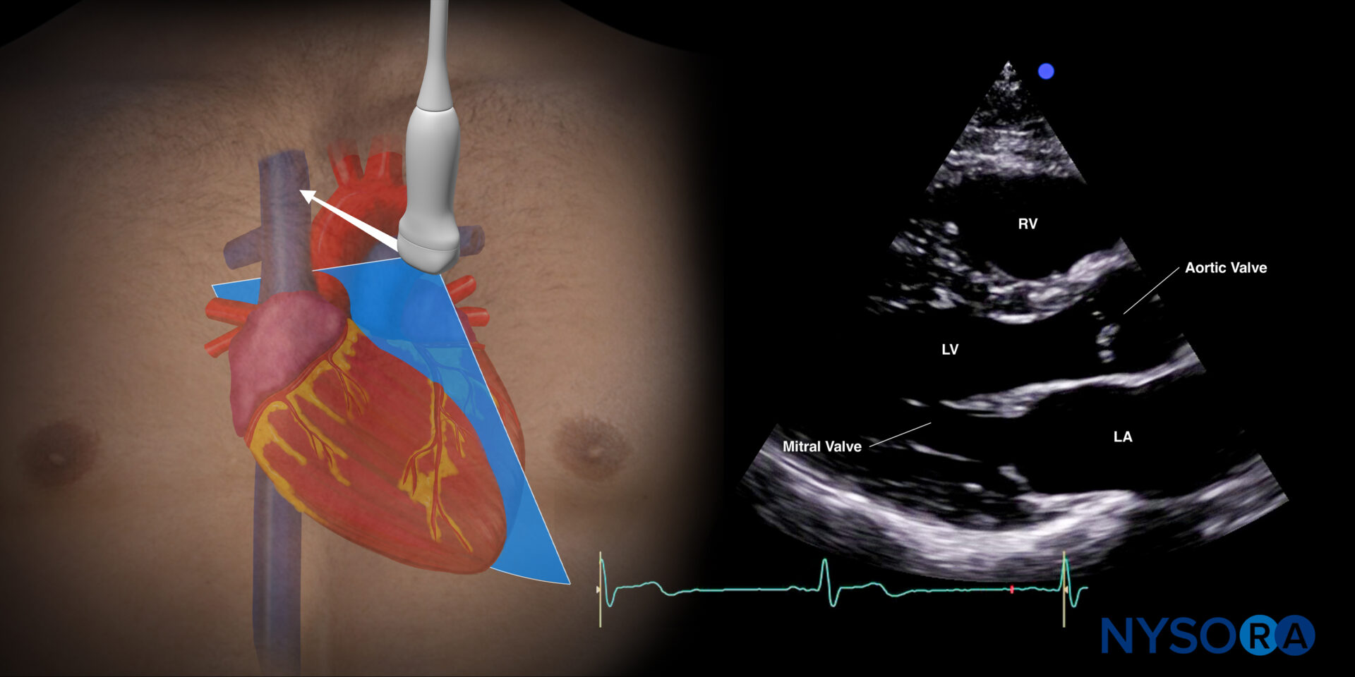
2. Parasternal long axis (PLAX) view
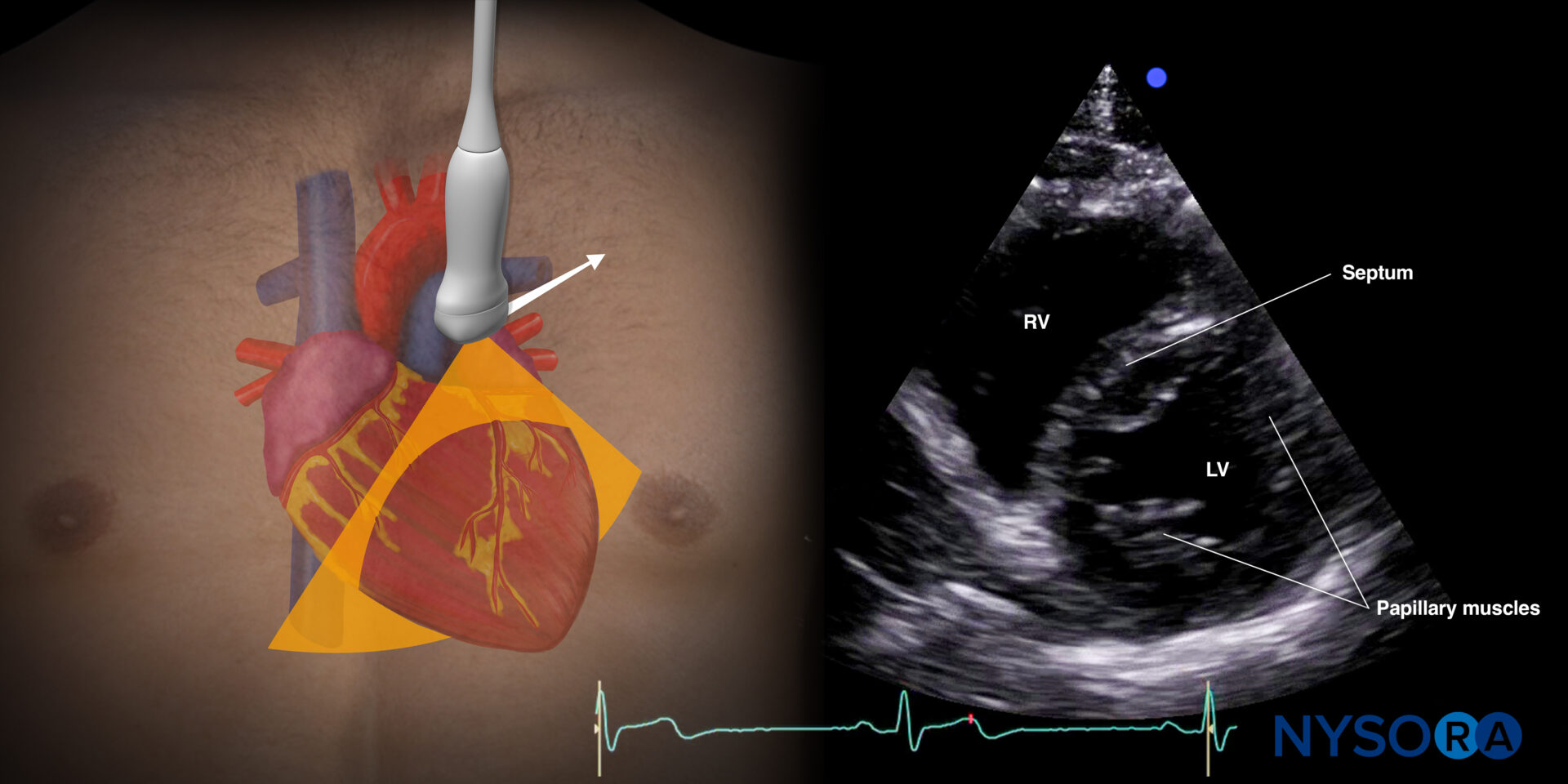
3. Parasternal short axis (PSAX) view
Each view offers specific insights into the movement and thickening of the myocardium during systole.
Qualitative assessment: The “eyeball” method
The eyeball method focuses on observing three key parameters to estimate left ventricular function:
- Inward movement of the endocardium during systole
- Normal function: The left ventricular walls contract inward symmetrically, and the endocardium moves toward the center of the ventricle.
- Reduced function: Poor inward movement and asymmetric or minimal endocardial motion.
- Views to Use: Apical Four-Chamber (A4C), Parasternal Long Axis (PLAX), Parasternal Short Axis (PSAX)
- Myocardial thickening during systole
- Normal function: Myocardial wall thickens by more than one-third during systole.
- Reduced function: Myocardial thickening is less than one-third, indicating impaired contraction.
- Views to Use: A4C, PLAX, PSAX
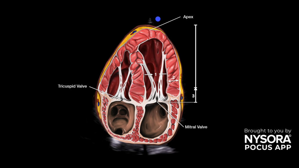
- Longitudinal shortening (mitral annulus to apex movement)
- Normal function: The base of the mitral valve (annulus) moves closer to the apex of the heart during systole by more than 1 cm.
- Reduced function: The shortening distance is less than 1 cm.
- Views to Use: A4C, PLAX
- Motion of the mitral valve toward the septum
- Normal function: The anterior mitral valve leaflet approximates well toward the septum; the distance is less than 1 cm.
- Reduced function: Poor leaflet motion with a distance greater than 1 cm.
- View to Use: Parasternal Long Axis (PLAX)
Steps to perform left ventricular assessment
Follow these steps during a POCUS evaluation:
- Start with apical four-chamber view:
- Focus on the inward movement of the ventricular walls and observe myocardial thickening.
- Evaluate longitudinal shortening by tracking the mitral annulus-to-apex movement.
- Move to parasternal long axis view:
- Assess wall motion and thickening.
- Observe mitral valve motion toward the septum.
- Switch to parasternal short axis view:
- Examine the symmetric inward movement of the ventricular walls.
- Evaluate myocardial thickening across the short axis.
- Utilize M-mode for E-Point septal separation (EPSS):
- Measure the distance between the anterior mitral valve leaflet and the septum during maximal excursion.
- EPSS > 1 cm indicates reduced systolic function.
Tips for reliable assessments
- Place a cursor or pointer in the center of the left ventricle to observe motion.
- In cases of myocardial infarction, endocardial excursion and thickening are affected first; diminished mitral valve motion appears later.
- Conditions like mitral stenosis, prosthetic valves, or hypertrophic septum can limit the reliability of this qualitative method.
Clinical Case Example
Patient: 71-year-old male with a history of coronary artery disease, chronic hypertension, and type 2 diabetes.
Presentation: Chest pain.
Assessment: POCUS was performed using the Apical four-chamber view.
- Observed inward wall motion, myocardial thickening, and longitudinal shortening indicated reduced left ventricular function.
Outcome: Findings guided early intervention and management in an acute setting.
Conclusion
Qualitative assessment of left ventricular function using POCUS is an invaluable bedside tool, particularly in emergency and critical care scenarios. Clinicians can quickly distinguish between normal and reduced ventricular function by focusing on wall motion, myocardial thickening, and mitral valve motion, facilitating prompt and appropriate management.
For an enhanced clinical experience, explore tools like NYSORA’s POCUS App to refine your skills and elevate patient care.
Download NYSORA’s POCUS App
Unleash the potential of POCUS and deliver exceptional care. Download HERE.