New POCUS Course: Airway Assessment!
We are thrilled to announce the launch of our Airway Assessment course, now available in the POCUS App! This comprehensive course is designed to equip healthcare professionals with the knowledge and skills to perform advanced airway management using ultrasound techniques.
Learning objectives
You will gain expertise in:
- Understanding the anatomy and physiology of the upper airway.
- Identifying key upper airway structures.
- mastering the scanning techniques to assess the upper airway
Why airway ultrasound?
- Safe and non-invasive: Quick and painless for patients.
- Highly accurate: Provides real-time assessment of airway structures and conditions.
- Improved outcomes: Reduces complications in difficult airway scenarios, a leading cause of anesthesia-related mortality.
- Evidence-based: Confirming ETT placement via ultrasound shows 98% sensitivity and specificity.
What you’ll learn
This course covers essential topics in airway ultrasound, including:
Upper airway anatomy and physiology
Gain a solid understanding of the functional anatomy.
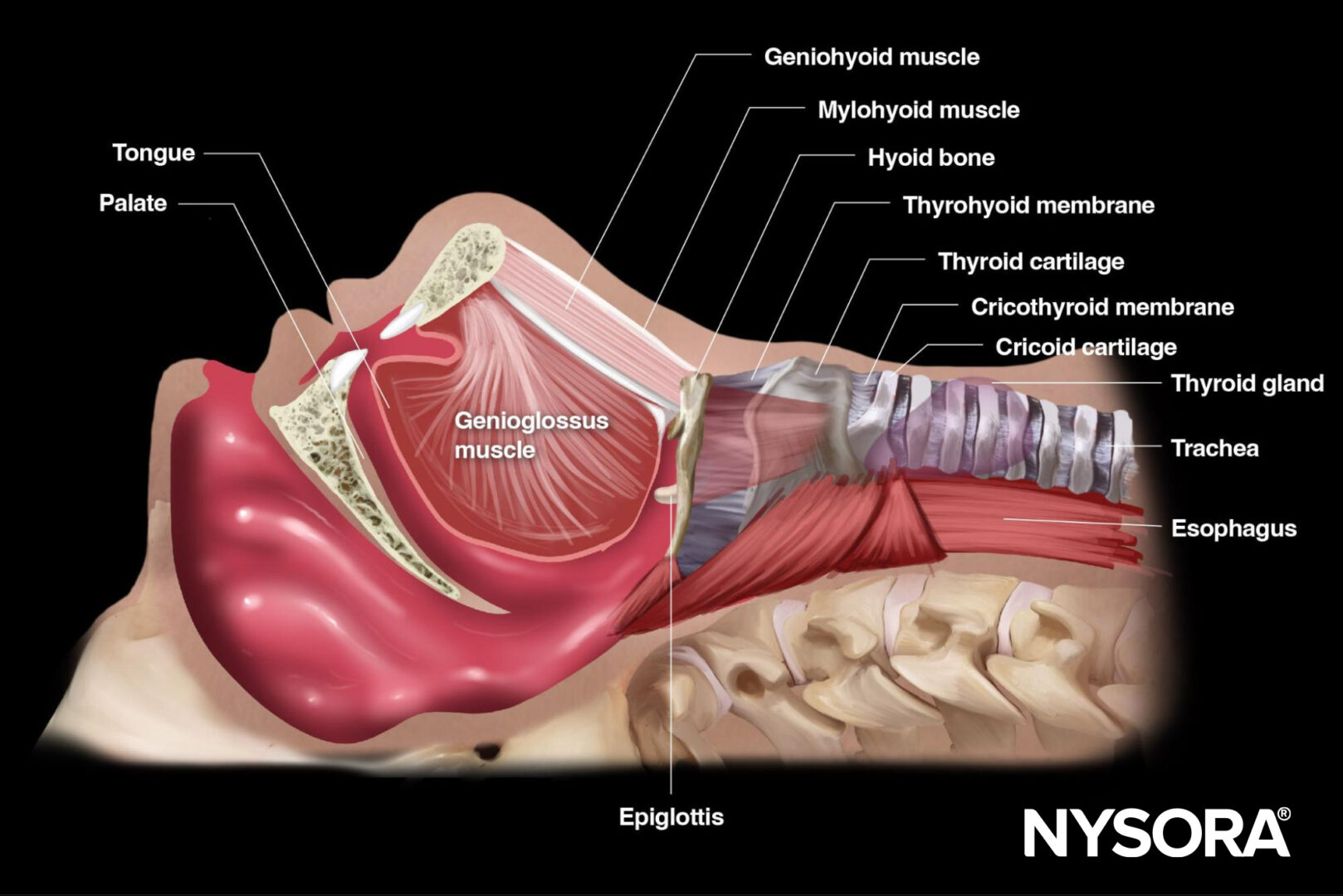
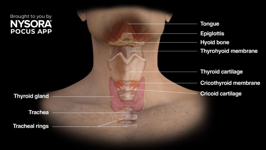
Identification of upper airway structures with ultrasound
Detailed descriptions help you identify structures clearly, for instance:
- Hyoid bone – recognizable on ultrasound as a linear hyperechoic inverted U-shaped structure with two greater horns (cornua).
- Thyroid cartilage – ultrasound appearance as a hypoechoic inverted V-shaped or triangular structure.
- Cricothyroid membrane – appears as a hyperechoic horizontal line.
Ultrasound imaging techniques
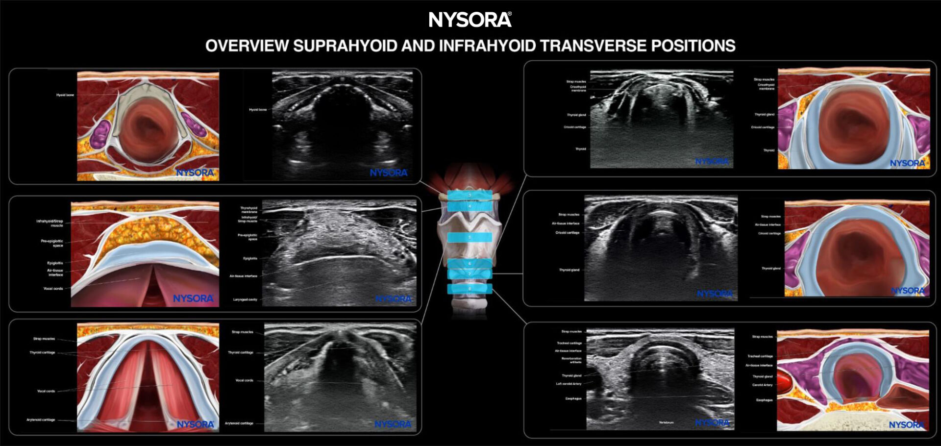
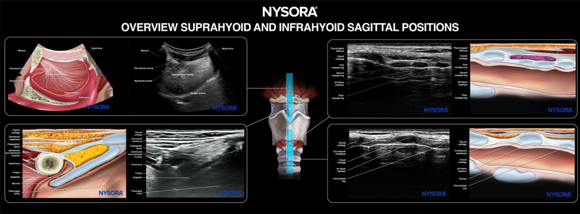
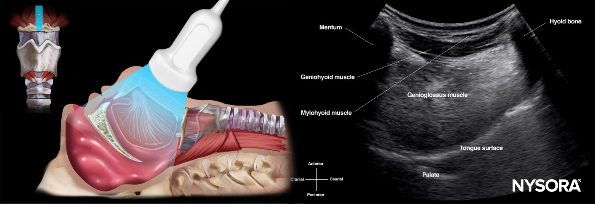
- Suprahyoid area
-
- Place the transducer longitudinally below the mandible and above the hyoid bone.
- Ultrasound image shows a fan-shaped tongue with a typical striated appearance.
- Underneath the mylohyoid and geniohyoid muscles, the tongue appears fan-shaped, with intrinsic muscles providing a striated appearance.
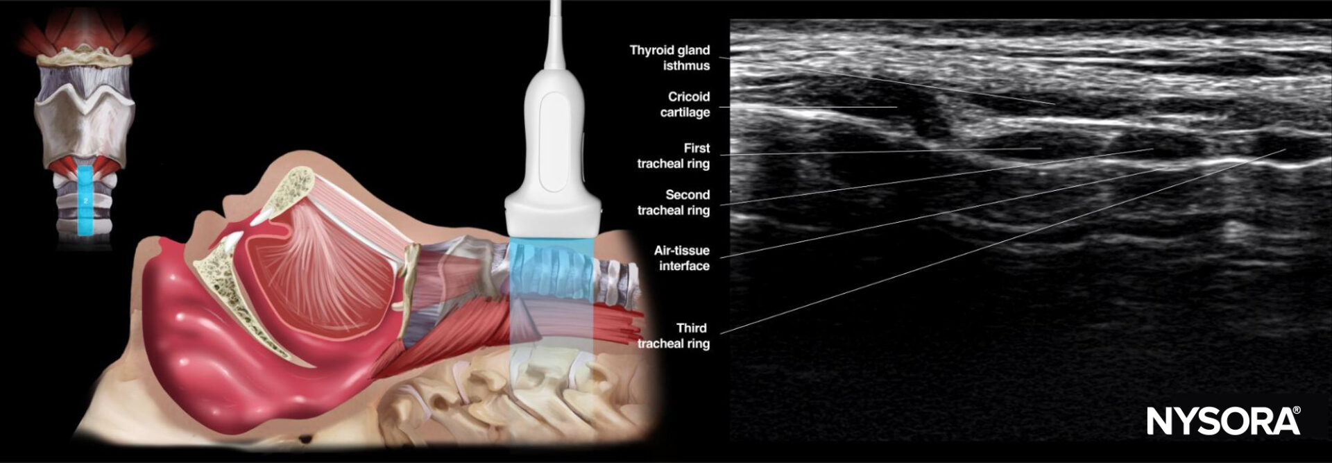
- Infrahyoid area: Sagittal ‘string of pearls’ technique
-
- Start from the trachea, sliding the probe cranially.
- The tracheal rings appear as dark hypoechoic structures, resembling a pearl necklace.
- Infrahyoid transverse approach
Position the transducer transversely on the midline of the neck. Scan cranially to caudally, visualizing key landmarks:
- Hyoid bone
- Thyrohyoid membrane
- Thyroid cartilage
- Cricothyroid membrane
- Cricoid cartilage
- Trachea

Who is this for?
This course is ideal for:
- Anesthesiologists.
- Emergency medicine practitioners.
- Critical care specialists.
- Surgeons and OR staff.
- Residents and medical students.
The Airway Assessment course is now live in the POCUS App. Download the POCUS App today and start your journey to mastering airway management!