Tips for the Thoracic Paravertebral Block (Transverse Scan)
The TPVB is indicated to provide anesthesia and analgesia of any hemithorax surgical procedure and when the associated pain is mainly unilateral of the chest and abdomen.
It is often used as an adjunct to multimodal postoperative analgesia in thoracic surgery, breast surgery, renal surgery, video-assisted thoracoscopic surgery, minimally invasive cardiac surgery, and more frequently indicated recently in breast reconstruction surgery in combination with general anesthesia.
Here at NYSORA, we follow these scanning tips when performing the Thoracic Paravertebral Block:
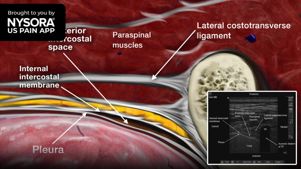
Place the transducer lateral to the spinous process with the orientation marker directed to the patient’s right side and identify the following structures on a transverse scan of the thoracic paravertebral region:
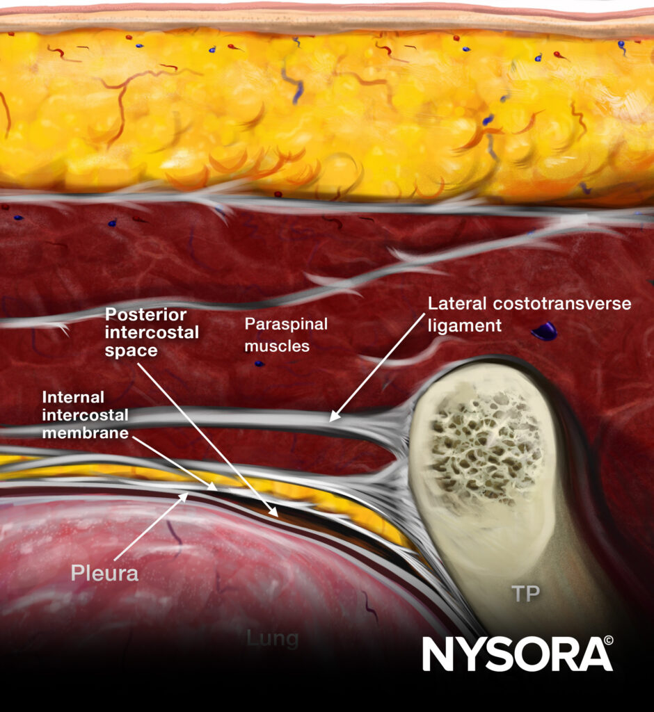
- Paraspinal muscles: Clearly delineated and lie superficial to the transverse processes.
- Transverse processes: Hyperechoic, anterior to which there is a dark acoustic shadow that completely obscures the thoracic paravertebral space (TPVS).
- Pleura: Hyperechoic, lateral to the transverse process, moves with respiration and exhibits the typical lung sliding sign.
Posterior intercostal space: Hypoechoic space between the parietal pleura and internal intercostal membrane, which is the medial extension of the internal intercostal muscle and is continuous medially with the superior costotransverse ligament. This space represents the medial limit of the posterior intercostal space or the apex of the TPVS.

Sonoanatomy

Reverse Ultrasound Anatomy
Download the US Pain App HERE to read other tips on managing acute and chronic pain and to access the complete guide to ultrasound-guided chronic pain blocks.


