Interventional Pain App - NYSORA

Explore NYSORA knowledge base for free:

Standardized Fluoroscopy-Guided Injections for Chronic Pain Management

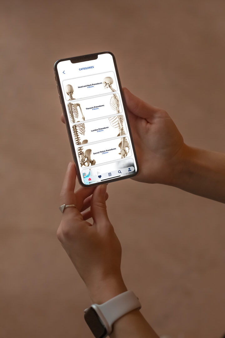
Complete learning material for your FIPP Exam on your mobile phone and tablet!

The single best resource for preparing for the FIPP examination that is also guaranteed to improve patient care and safety standards!
Highly practical mnemonics to help you memorize structures important for success, and avoid complications!
Join the community of over 30,000 professionals worldwide and see for yourself why interventional pain management app is the first and the most authoritative guide on fluoroscopy-guided injections.
20+ Standard, Highly Practical Chronic Pain Interventions
Gain access to the most authoritative guide on fluoroscopy-guided injections: Interlaminar Cervical Epidural Injection, Intra Articular Cervical Facet Joint Block, C2-T1 – Posterior and Lateral Approach, Intercostal Nerve Block, Sacroiliac Joint Injection, Sacroiliac Joint Radiofrequency Ablation (Bipolar Palisade Technique), Superior Hypogastric Plexus Block – Anterior Approach, Neuroplasty (Caudal, Transgrade and Transforaminal approach), Superior Hypogastric Plexus Block – Transdiscal Approach, Splanchnic Block and Radiofrequency Ablation
Choose between monthly and annual
Subscribe and get full access! With our annual subscription, subscribers will receive proof of payment for education reimbursement. You also save 15% if you choose the annual subscription instead of monthly!
Annually
Monthly
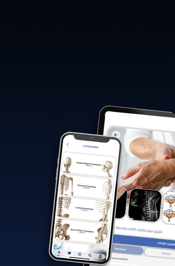
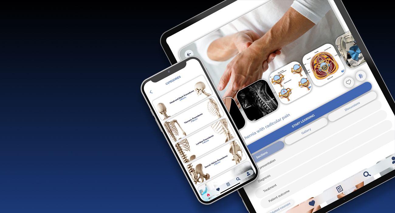
Pain Rx App that is Optimized for Mobile phones and Tablets
The most authoritative guide on standardized principles and techniques of interventional chronic pain interventions, including joint injections, nerve blocks, radiofrequency ablations, stimulation procedures, and more.

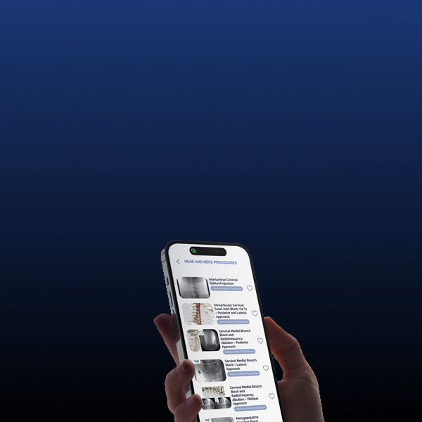
Extensive Image Gallery​

News from the app

Hidden risks of gadolinium-based contrast agents in interventional pain medicine

A recent study by Hallo-Carrasco et al. highlights the potential dangers of gadolinium-based contrast media (GCM) in interventional pain medicine. This research, published in Regional Anesthesia & Pain Medicine (2024), investigates adverse events linked to using GCM during spine procedures where inadvertent intrathecal administration is a risk. Here’s a comprehensive look at the findings, implications, and best practices surrounding GCM use in these procedures. Overview of gadolinium-based contrast media (GCM) Gadolinium-based contrast agents are an alternative for patients with documented allergies to iodinated contrast media (ICM). While traditionally safer for imaging, GCM carries specific risks: Toxicity risks: Gadolinium ions (Gd3+) are inherently toxic; chelating agents stabilize the compound and reduce these risks. Neurotoxicity: When inadvertently injected into the intrathecal (spinal) space, GCM can cause severe neurotoxic effects. Brain retention: There are concerns about gadolinium deposits in brain tissues following repeated exposure. Key findings  Conducted via a retrospective review of medical records, the study investigated 508 patients who received GCM for spine-related procedures between 2019 and 2022. Here’s what was uncovered: Adverse event rate: 23 patients (3.3%) experienced adverse events potentially related to GCM. Common symptoms included severe pain, dizziness, headache, and, in one case, multifocal stroke. Patient demographics: A significant majority of patients were white females with a mean age of 67.55 years. Indications for GCM use: The predominant reason for using GCM was a documented iodine-related allergy. However, only 1% of these cases involved high-risk allergy reactions that could justify substituting ICM. Severity of adverse events: While most were manageable, some cases required hospitalization. The study documented one severe incident of stroke following GCM administration. Adverse reactions and symptoms Adverse effects following inadvertent intrathecal injection of GCM included: Severe pain: Reported within days of the procedure, sometimes requiring hospitalization. Headaches: Particularly postural headaches, which align with findings from other […]

December 3, 2024

Case Study: Cervical Hernia with Radicular Pain

Cervical disc herniation is a common condition that can cause significant pain, particularly when it leads to radicular pain. This article provides an in-depth look at a case of cervical hernia with radicular pain, detailing the diagnosis, treatment, and outcome. The focus will be on the Percutaneous Laser Disc Decompression (PLDD) procedure, a minimally invasive treatment option for patients with disc herniation who have not responded to conservative therapy. Case presentation A 41-year-old woman presented with cervical pain radiating to her left upper limb. The pain was aggravated by physical activities, including CrossFit, and was described as persistent, with shock-like sensations, pins and needles, and burning feelings. Pain intensity: Baseline pain was measured at 4/10 on the Numerical Rating Scale (NRS), peaking at 8/10 during episodes of exertion. Neurological evaluation: A detailed physical examination confirmed radicular pain following the C7 nerve root dermatome. Diagnosis Medical history and symptoms The patient reported no prior medical conditions but engaged in frequent high-intensity exercises, which may have contributed to the worsening of symptoms. The pain was consistent with radiculopathy, a condition characterized by nerve irritation due to a herniated disc. Physical examination findings: The Spurling test, a diagnostic maneuver used to assess cervical nerve root compression, was positive. The test aggravated the patient’s neck pain and reproduced the arm pain, indicating C7 nerve root involvement. Imaging: Magnetic Resonance Imaging (MRI) revealed a C6-C7 posterior disc protrusion with mild left-sided uncovertebral arthrosis, moderately narrowing the neural foramen. Final diagnosis The patient was diagnosed with cervical radiculopathy, specifically brachial plexus root pain caused by the C6-C7 disc herniation. This type of pain results from compression or irritation of the cervical nerve roots, often leading to pain that radiates down the arm. Treatment: Percutaneous Laser Disc Decompression (PLDD) What is PLDD? PLDD is a minimally invasive […]

February 22, 2024

Case study: Cancer-related lower abdominal and pelvic pain

Cancer-related chronic pain is a debilitating condition that can significantly impact a patient’s quality of life. In this article, we will explore the case of a 55-year-old woman with cervical uterine malignancy and severe lower abdominal and pelvic pain. We will focus on her diagnosis, pain management, and the use of a superior hypogastric plexus neurolytic block to alleviate her chronic pain. This procedure offers a targeted approach for managing pain associated with pelvic malignancies. Case presentation A 55-year-old woman presented with persistent lower abdominal and pelvic pain. She had been diagnosed with moderately differentiated non-keratinizing squamous cell carcinoma of the uterine cervix six months prior. Despite receiving extensive chemotherapy and radiotherapy, her pain had escalated, becoming a significant challenge in her treatment journey. Diagnosis: Cervical uterine malignancy. Previous treatments: 5 sessions of chemotherapy 20 sessions of radiotherapy Pain characteristics Type: Severe, constant, with episodes of shooting, stabbing, and pressure-like sensations. Location: Lower abdomen and pelvis. Intensity: Baseline pain was rated at 7-8/10 on the Numerical Rating Scale (NRS), with peaks reaching 10/10 during episodes of excruciating pain. Duration: Episodes lasting for several seconds occurred multiple times per day, severely impacting her quality of life. Current pain management The patient’s pain was being managed with a multimodal approach, including: Gabapentin: 600 mg orally every eight hours. Duloxetine: 60 mg orally once daily. Methadone: 5 mg orally every eight hours. Paracetamol: 1000 mg orally every eight hours. Despite this regimen, her pain remained challenging to control, particularly the breakthrough episodes, which were unresponsive to conventional pain medications. Physical examination and imaging Physical exam: Revealed deep tenderness in the lower abdomen with organomegaly. Neurological examination of the legs was normal. MRI findings: Showed a large mass in the lower uterine cervix and upper vagina, with additional masses identified in the iliac, para-aortic, […]

February 8, 2024

Case study: Upper limb complex regional pain syndrome (CRPS)

Case study: Upper limb complex regional pain syndrome (CRPS) Complex Regional Pain Syndrome (CRPS) is a challenging and often debilitating condition characterized by prolonged pain and inflammation following injury. This case study highlights a multidisciplinary approach to diagnosing and managing CRPS in a patient with a history of trauma. Focusing on the innovative use of stellate ganglion blocks and an oblique fluoroscopic approach, this article provides an in-depth understanding of the techniques and outcomes. Case presentation Patient profile: Age: 59 years Gender: Male History: Left wrist fracture following a traffic accident, complicated by infection and neuropathic pain. Key medical history Comorbidities: Coeliac disease. Injury: Left wrist fracture treated with surgical fixation. Complications: Wound infection requiring debridement and persistent neuropathic pain. Symptoms Persistent left-hand pain radiating to the forearm and arm. Pain exacerbation with intermittent swelling and discoloration. Neuropathic pain with allodynia (pain from non-painful stimuli). Pain scores: Baseline NRS 5, peaking at 8. Physical examination Mild trophic changes and erythema of the left hand. No dystonia or overt motor deficits. Blood tests: No leukocytosis or elevated inflammatory markers. Diagnosis Complex Regional Pain Syndrome (CRPS) of the left upper limb. Key diagnostic features (Budapest criteria): Persistent pain disproportionate to the initial injury. Sensory changes: Allodynia and hyperalgesia. Vasomotor dysfunction: Intermittent swelling and discoloration. Exclusion of alternative diagnoses via clinical evaluation and imaging. Understanding CRPS What is CRPS? CRPS is a chronic pain condition typically triggered by trauma or nerve injury. It involves: Excessive pain and inflammation. Changes in skin color, temperature, and texture. Joint stiffness and impaired mobility. Pathophysiology The exact cause of CRPS is unclear but may involve: Peripheral and central nervous system sensitization. Dysregulated inflammatory responses. Treatment overview A multidisciplinary approach is crucial for managing CRPS. Pharmacological management Pregabalin: 75 mg twice daily for neuropathic pain. Amitriptyline: 25 mg […]

December 28, 2023

Interventional Pain App Explained

We are excited to share thrilling news with our community! NYSORA launches a new INTERVENTIONAL PAIN mobile app! With state-of-the-art, highly practical material from Springer’s best selling book “Interventional Pain: A Step-by-Step Guide for the FIPP Exam”, it’s a must-have for practitioners focusing on fluoroscopy-guided pain procedures. Read more about this great app here: https://www.nysora.com/nysora-interventional-pain-app/! The image-driven interventional pain procedures APP is created by the key opinion leaders with years of experience. With this app you will have instantaneous access to: Interventional Pain Procedures on the GO; Standardized Fluoroscopy-Guided Interventional Pain Procedures; Complete learning material for FIPP Exam on your mobile; Evidence-based recommendations asked at FIPP Exam; Tips on how to obtain the best images.. and much more! Download NYSORA’s Interventional Pain App on the buttons down below and start with your 7 days free trial!

July 22, 2021

NEW RELEASE: Mobile Guide to Fluoroscopy Interventional Pain Management!

NYSORA launches NEW Interventional Pain App by Springer, published by the world’s top experts on Interventional Chronic Pain management. We are excited to share thrilling news with our community! NYSORA has launched a new mobile app – Interventional Pain on July 19th, 2021. This state of the art, highly practical material from Springer’s best selling book “Interventional Pain : A Step-by-Step Guide for the FIPP Exam” by Springer Nature, is a must-have for practitioners with focus on fluoroscopy-guided pain procedures.  Why should you get this APP? The image-driven interventional pain procedures APP is created by the key opinion leaders with years of experience. With this app you will have instantaneous access to: Interventional Pain Procedures on the GO; Standardized Fluoroscopy-Guided Interventional Pain Procedures; Complete learning material for FIPP Exam on your mobile; Evidence-based recommendations asked at FIPP Exam; Tips on how to obtain best images; Amazing mnemonics for remembering tips for success and complications;  Best resource for both examinees and examiners; Improves patient care and safety; Fluoroscopic guidance for everything from cervical spine interventions, to sacroiliac and facet injections, to radiofrequency ablations. The newest edition, thoroughly updated with latest recommendations that cover the entire therapeutic area applicable to Fluoroscopic-guided Interventional Pain Medicine.  NYSORA’s lead, Dr. Hadzic points out that this app is the single best resource featuring all standardized and evidence-based interventions in the practice of interventional pain medicine. Importantly, the app features clear guidance to diagnostic and therapeutic evidence-based recommendations, and it is the single best resource for preparing for the FIPP examination, administered by the World Institute of Pain (WIP).  “We are committed to continually expand the educational resources utilizing our 20 years experience of transferring knowledge. Our educational mobile learning platforms benefit thousands of students and professionals who use NYSORA apps. For this new APP, we listened […]

July 19, 2021

Hidden risks of gadolinium-based contrast agents in interventional pain medicine

A recent study by Hallo-Carrasco et al. highlights the potential dangers of gadolinium-based contrast media (GCM) in interventional pain medicine. This research, published in Regional Anesthesia & Pain Medicine (2024), investigates adverse events linked to using GCM during spine procedures where inadvertent intrathecal administration is a risk. Here’s a comprehensive look at the findings, implications, and best practices surrounding GCM use in these procedures. Overview of gadolinium-based contrast media (GCM) Gadolinium-based contrast agents are an alternative for patients with documented allergies to iodinated contrast media (ICM). While traditionally safer for imaging, GCM carries specific risks: Toxicity risks: Gadolinium ions (Gd3+) are inherently toxic; chelating agents stabilize the compound and reduce these risks. Neurotoxicity: When inadvertently injected into the intrathecal (spinal) space, GCM can cause severe neurotoxic effects. Brain retention: There are concerns about gadolinium deposits in brain tissues following repeated exposure. Key findings  Conducted via a retrospective review of medical records, the study investigated 508 patients who received GCM for spine-related procedures between 2019 and 2022. Here’s what was uncovered: Adverse event rate: 23 patients (3.3%) experienced adverse events potentially related to GCM. Common symptoms included severe pain, dizziness, headache, and, in one case, multifocal stroke. Patient demographics: A significant majority of patients were white females with a mean age of 67.55 years. Indications for GCM use: The predominant reason for using GCM was a documented iodine-related allergy. However, only 1% of these cases involved high-risk allergy reactions that could justify substituting ICM. Severity of adverse events: While most were manageable, some cases required hospitalization. The study documented one severe incident of stroke following GCM administration. Adverse reactions and symptoms Adverse effects following inadvertent intrathecal injection of GCM included: Severe pain: Reported within days of the procedure, sometimes requiring hospitalization. Headaches: Particularly postural headaches, which align with findings from other […]

December 3, 2024

Case Study: Cervical Hernia with Radicular Pain

Cervical disc herniation is a common condition that can cause significant pain, particularly when it leads to radicular pain. This article provides an in-depth look at a case of cervical hernia with radicular pain, detailing the diagnosis, treatment, and outcome. The focus will be on the Percutaneous Laser Disc Decompression (PLDD) procedure, a minimally invasive treatment option for patients with disc herniation who have not responded to conservative therapy. Case presentation A 41-year-old woman presented with cervical pain radiating to her left upper limb. The pain was aggravated by physical activities, including CrossFit, and was described as persistent, with shock-like sensations, pins and needles, and burning feelings. Pain intensity: Baseline pain was measured at 4/10 on the Numerical Rating Scale (NRS), peaking at 8/10 during episodes of exertion. Neurological evaluation: A detailed physical examination confirmed radicular pain following the C7 nerve root dermatome. Diagnosis Medical history and symptoms The patient reported no prior medical conditions but engaged in frequent high-intensity exercises, which may have contributed to the worsening of symptoms. The pain was consistent with radiculopathy, a condition characterized by nerve irritation due to a herniated disc. Physical examination findings: The Spurling test, a diagnostic maneuver used to assess cervical nerve root compression, was positive. The test aggravated the patient’s neck pain and reproduced the arm pain, indicating C7 nerve root involvement. Imaging: Magnetic Resonance Imaging (MRI) revealed a C6-C7 posterior disc protrusion with mild left-sided uncovertebral arthrosis, moderately narrowing the neural foramen. Final diagnosis The patient was diagnosed with cervical radiculopathy, specifically brachial plexus root pain caused by the C6-C7 disc herniation. This type of pain results from compression or irritation of the cervical nerve roots, often leading to pain that radiates down the arm. Treatment: Percutaneous Laser Disc Decompression (PLDD) What is PLDD? PLDD is a minimally invasive […]
February 22, 2024

Case study: Cancer-related lower abdominal and pelvic pain

Cancer-related chronic pain is a debilitating condition that can significantly impact a patient’s quality of life. In this article, we will explore the case of a 55-year-old woman with cervical uterine malignancy and severe lower abdominal and pelvic pain. We will focus on her diagnosis, pain management, and the use of a superior hypogastric plexus neurolytic block to alleviate her chronic pain. This procedure offers a targeted approach for managing pain associated with pelvic malignancies. Case presentation A 55-year-old woman presented with persistent lower abdominal and pelvic pain. She had been diagnosed with moderately differentiated non-keratinizing squamous cell carcinoma of the uterine cervix six months prior. Despite receiving extensive chemotherapy and radiotherapy, her pain had escalated, becoming a significant challenge in her treatment journey. Diagnosis: Cervical uterine malignancy. Previous treatments: 5 sessions of chemotherapy 20 sessions of radiotherapy Pain characteristics Type: Severe, constant, with episodes of shooting, stabbing, and pressure-like sensations. Location: Lower abdomen and pelvis. Intensity: Baseline pain was rated at 7-8/10 on the Numerical Rating Scale (NRS), with peaks reaching 10/10 during episodes of excruciating pain. Duration: Episodes lasting for several seconds occurred multiple times per day, severely impacting her quality of life. Current pain management The patient’s pain was being managed with a multimodal approach, including: Gabapentin: 600 mg orally every eight hours. Duloxetine: 60 mg orally once daily. Methadone: 5 mg orally every eight hours. Paracetamol: 1000 mg orally every eight hours. Despite this regimen, her pain remained challenging to control, particularly the breakthrough episodes, which were unresponsive to conventional pain medications. Physical examination and imaging Physical exam: Revealed deep tenderness in the lower abdomen with organomegaly. Neurological examination of the legs was normal. MRI findings: Showed a large mass in the lower uterine cervix and upper vagina, with additional masses identified in the iliac, para-aortic, […]

February 8, 2024

Case study: Upper limb complex regional pain syndrome (CRPS)

Case study: Upper limb complex regional pain syndrome (CRPS) Complex Regional Pain Syndrome (CRPS) is a challenging and often debilitating condition characterized by prolonged pain and inflammation following injury. This case study highlights a multidisciplinary approach to diagnosing and managing CRPS in a patient with a history of trauma. Focusing on the innovative use of stellate ganglion blocks and an oblique fluoroscopic approach, this article provides an in-depth understanding of the techniques and outcomes. Case presentation Patient profile: Age: 59 years Gender: Male History: Left wrist fracture following a traffic accident, complicated by infection and neuropathic pain. Key medical history Comorbidities: Coeliac disease. Injury: Left wrist fracture treated with surgical fixation. Complications: Wound infection requiring debridement and persistent neuropathic pain. Symptoms Persistent left-hand pain radiating to the forearm and arm. Pain exacerbation with intermittent swelling and discoloration. Neuropathic pain with allodynia (pain from non-painful stimuli). Pain scores: Baseline NRS 5, peaking at 8. Physical examination Mild trophic changes and erythema of the left hand. No dystonia or overt motor deficits. Blood tests: No leukocytosis or elevated inflammatory markers. Diagnosis Complex Regional Pain Syndrome (CRPS) of the left upper limb. Key diagnostic features (Budapest criteria): Persistent pain disproportionate to the initial injury. Sensory changes: Allodynia and hyperalgesia. Vasomotor dysfunction: Intermittent swelling and discoloration. Exclusion of alternative diagnoses via clinical evaluation and imaging. Understanding CRPS What is CRPS? CRPS is a chronic pain condition typically triggered by trauma or nerve injury. It involves: Excessive pain and inflammation. Changes in skin color, temperature, and texture. Joint stiffness and impaired mobility. Pathophysiology The exact cause of CRPS is unclear but may involve: Peripheral and central nervous system sensitization. Dysregulated inflammatory responses. Treatment overview A multidisciplinary approach is crucial for managing CRPS. Pharmacological management Pregabalin: 75 mg twice daily for neuropathic pain. Amitriptyline: 25 mg […]

December 28, 2023

Interventional Pain App Explained

We are excited to share thrilling news with our community! NYSORA launches a new INTERVENTIONAL PAIN mobile app! With state-of-the-art, highly practical material from Springer’s best selling book “Interventional Pain: A Step-by-Step Guide for the FIPP Exam”, it’s a must-have for practitioners focusing on fluoroscopy-guided pain procedures. Read more about this great app here: https://www.nysora.com/nysora-interventional-pain-app/! The image-driven interventional pain procedures APP is created by the key opinion leaders with years of experience. With this app you will have instantaneous access to: Interventional Pain Procedures on the GO; Standardized Fluoroscopy-Guided Interventional Pain Procedures; Complete learning material for FIPP Exam on your mobile; Evidence-based recommendations asked at FIPP Exam; Tips on how to obtain the best images.. and much more! Download NYSORA’s Interventional Pain App on the buttons down below and start with your 7 days free trial!

July 22, 2021

NEW RELEASE: Mobile Guide to Fluoroscopy Interventional Pain Management!

NYSORA launches NEW Interventional Pain App by Springer, published by the world’s top experts on Interventional Chronic Pain management. We are excited to share thrilling news with our community! NYSORA has launched a new mobile app – Interventional Pain on July 19th, 2021. This state of the art, highly practical material from Springer’s best selling book “Interventional Pain : A Step-by-Step Guide for the FIPP Exam” by Springer Nature, is a must-have for practitioners with focus on fluoroscopy-guided pain procedures.  Why should you get this APP? The image-driven interventional pain procedures APP is created by the key opinion leaders with years of experience. With this app you will have instantaneous access to: Interventional Pain Procedures on the GO; Standardized Fluoroscopy-Guided Interventional Pain Procedures; Complete learning material for FIPP Exam on your mobile; Evidence-based recommendations asked at FIPP Exam; Tips on how to obtain best images; Amazing mnemonics for remembering tips for success and complications;  Best resource for both examinees and examiners; Improves patient care and safety; Fluoroscopic guidance for everything from cervical spine interventions, to sacroiliac and facet injections, to radiofrequency ablations. The newest edition, thoroughly updated with latest recommendations that cover the entire therapeutic area applicable to Fluoroscopic-guided Interventional Pain Medicine.  NYSORA’s lead, Dr. Hadzic points out that this app is the single best resource featuring all standardized and evidence-based interventions in the practice of interventional pain medicine. Importantly, the app features clear guidance to diagnostic and therapeutic evidence-based recommendations, and it is the single best resource for preparing for the FIPP examination, administered by the World Institute of Pain (WIP).  “We are committed to continually expand the educational resources utilizing our 20 years experience of transferring knowledge. Our educational mobile learning platforms benefit thousands of students and professionals who use NYSORA apps. For this new APP, we listened […]

July 19, 2021
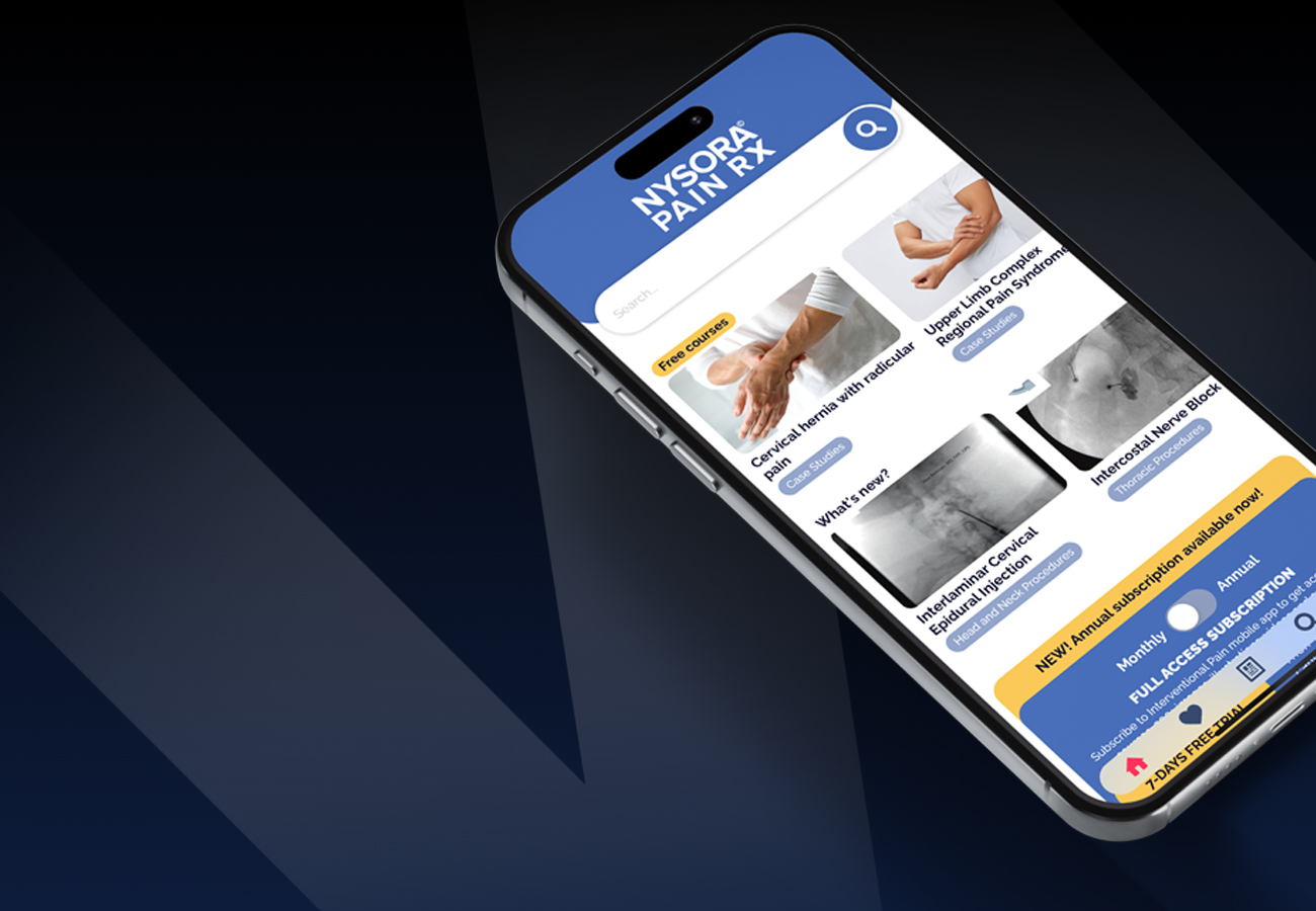
Pain Management App that is Optimized for Mobile phones and Tablets
35,000+
Downloads
Provides a timely and much-needed standardized approach to the 20 procedures tested at the FIPP exam
Based on the bestselling book by Springer Nature, Interventional Pain: A Step-by-Step Guide for the FIPP Exam, this app serves as a review for the Fellow of Interventional Pain Practice (FIPP) exam and functions as a concise guide for all interventional pain doctors.

Download Interventional Pain App Today and Get Started With a 7-day Free Trial

Frequently asked questions

Interventional pain refers to a specialized branch of medicine focused on diagnosing and treating pain through minimally invasive procedures. These procedures target the source of pain, providing relief and improving patients’ quality of life.

While interventional pain management is a crucial component of pain management, they are not exactly the same. Pain management encompasses various approaches to alleviate pain, including medications, physical therapy, lifestyle modifications, and interventional procedures. Interventional pain management specifically involves procedures such as nerve blocks, epidural injections, and spinal cord stimulation to address pain at its source.

The role of interventional pain management is to accurately diagnose and treat pain by targeting specific nerves, muscles, or joints with minimally invasive procedures. These techniques aim to reduce pain, improve function, and enhance the overall quality of life for patients suffering from chronic or acute pain conditions.

Non-interventional pain refers to pain management strategies that do not involve invasive procedures or interventions. This approach may include medications, physical therapy, acupuncture, chiropractic care, and psychological therapies aimed at managing pain without directly targeting the source through procedures like injections or nerve blocks.

Interventional pain management has evolved significantly over the years, with roots tracing back to the development of regional anesthesia techniques in the early 20th century. The field saw rapid advancement in the latter half of the century, particularly with the introduction of fluoroscopy and ultrasound-guided procedures. Today, interventional pain management continues to innovate with advancements in technology, techniques, and research, offering patients effective alternatives for pain relief.