Case study: Detecting intracranial hypertension using transcranial doppler ultrasound
Transcranial Doppler (TCD) ultrasound is a non-invasive tool utilized in point-of-care ultrasound (POCUS) for assessing cerebral blood flow dynamics. This case study explores the application of TCD in detecting intracranial hypertension in a clinical setting.
Case presentation:
- A 45-year-old male presented to the emergency department with severe headache, nausea, and blurred vision.
- Medical history included hypertension and a recent head trauma from a minor car accident.
Physical Examination:
- The patient was conscious but exhibited signs of increased intracranial pressure (ICP), such as papilledema and bradycardia.
- Initial neurological assessment showed no focal deficits.
Clinical Decision:
- Given the suspicion of intracranial hypertension, a TCD ultrasound was performed at the bedside to quickly assess cerebral blood flow dynamics and evaluate for raised ICP.
Indications for TCD
- Intracranial hypertension
- Suspected diagnosis of cerebral circulatory arrest
- Vasospasm detection
- Identification of midline shift
Essential Information on TCD
- TCD offers real-time information and can be performed at the bedside.
- It is not a replacement for CT scans but provides trending capabilities and immediate data.
Functional Anatomy and Machine Setup
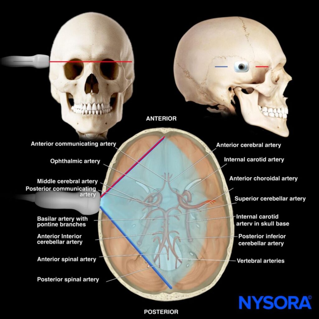
Anatomy:
- Key structures include the circle of Willis and intracranial arteries.
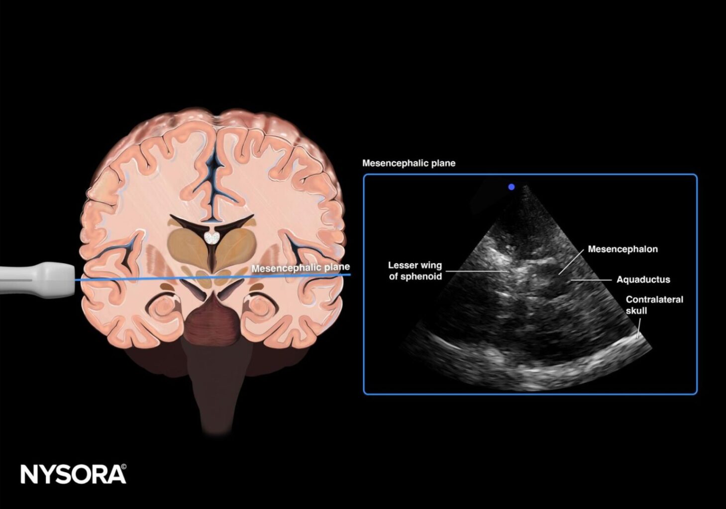
- The mesencephalic plane is critical for vascular assessment.
Machine Setup:
- Transducer: Phased array
- Preset: Transcranial (or cardiac)
- Orientation: Index marker toward the frontal bone/orbital
- Depth: 15 cm
Patient Positioning:
- Patient positioned supine with the head of the bed elevated to 30 degrees.
- Landmarks include the ear and temporomandibular joint.
- Transducer placed 2-3 cm above the temporomandibular joint at the level of the temporal bone.
Scanning Plane:
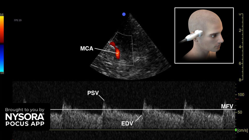
- Mesencephalic plane: Visualizes the middle cerebral artery (MCA) with red flow toward the transducer. Use pulsed wave Doppler to measure cerebral blood flow velocities.
Assessment Using TCD
- Pulsatility Index (PI): Calculated using the formula:
PI = (PSV – EDV)/ MFV
Where PSV is peak systolic velocity, EDV is end diastolic velocity, and MFV is mean flow velocity.
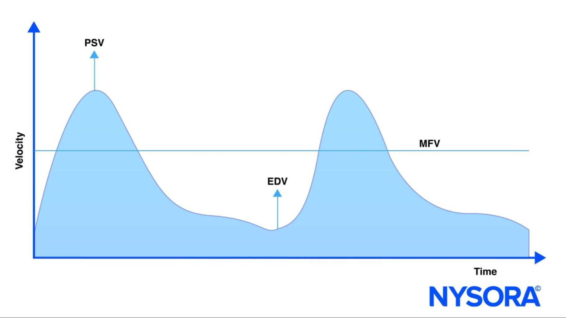
- A normal PI ranges from 0.5 to 1. A PI > 1.2 indicates increased ICP. The estimated ICP approximates PI x 10.
Findings:
- The patient’s PI was 1.4, suggesting raised ICP.
- Immediate intervention with measures to reduce ICP was initiated.
Conclusion
Transcranial Doppler is a valuable, non-invasive technique for assessing intracranial hypertension. By understanding cerebral anatomy and utilizing proper ultrasound techniques, healthcare providers can make rapid and accurate diagnoses, improving patient outcomes.
For more in-depth information on TCD and advanced applications, consider downloading NYSORA’s POCUS App for detailed resources, illustration, animations, and more.