Certain basic principles of optimizing an ultrasound image are applicable to all nerve blocks. For instance, sonography requires an understanding of mechanics and ergonomics. Novices are subject to errors such as probe fatigue, reversing probe orientation, and inadequate equipment preparation. To optimize the ultrasound image, the mnemonic PART (Pressure, Alignment, Rotation, Tilting) has been recommended.
Correct Pressure application can considerably improve the image quality; it is necessary to minimize the distance to the target and compress the underlying subcutaneous adipose tissues.
Alignment refers to placing the transducer in a position over the extremity (or trunk) at which the underlying nerve is optimally located for needle advancement.
Rotation allows adjusting the view of the structures achieving a true axial or longitudinal view of the target.
Tilting helps to bring the face of the probe into a perpendicular arrangement with the underlying target to maximize the number of returning echoes and thus provide the best image.
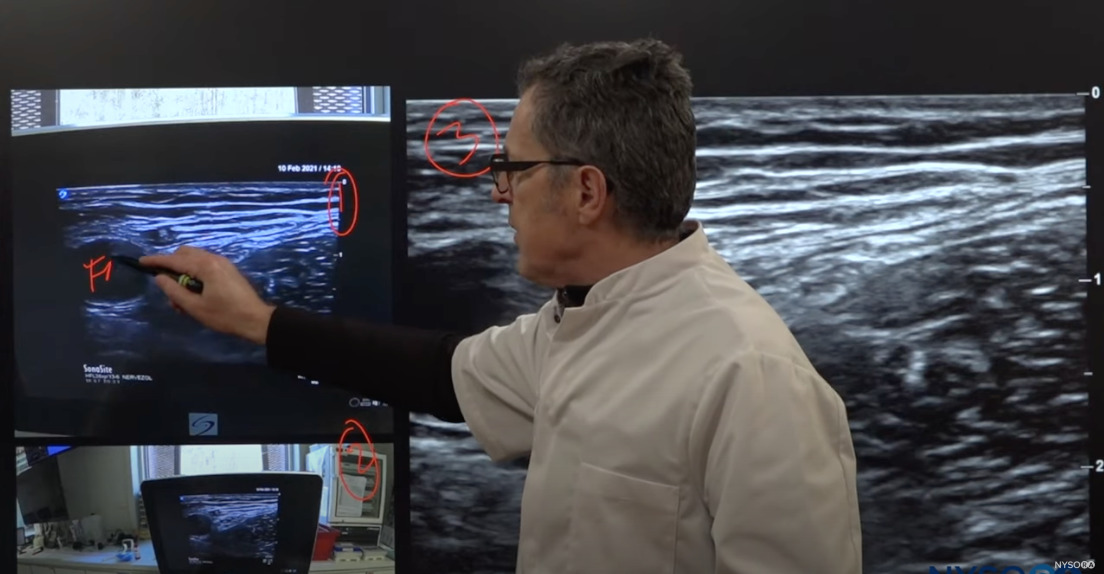
The following video demonstrates how using NYSORA SIMULATORS™ is the best way to learn the essential ultrasound maneuvres, hand-eye-coordination, and anatomy recognition.
Additional features from the essential ultrasound trainer:
- Allows needle-target practice in-plane and out-of-plane
- Facilitates acquisition of hand-eye coordination
- Teaches essential ultrasound maneuvres
- Allows practice ultrasound-guided vascular access
For more information on NYSORA SIMULATORS™, please click here.


