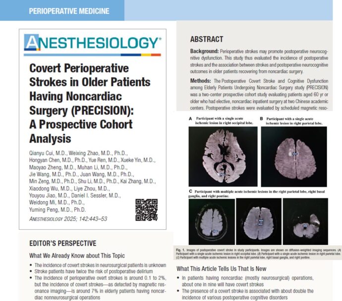
Ultrasound-guided single popliteal sciatic nerve block for postoperative analgesia in calcaneal fractures
The popliteal sciatic nerve block is indicated for foot and ankle surgery, foot and toe amputation, and Achilles tendon surgery. It targets the sciatic nerve at or slightly above its division into the tibial and common peroneal nerves. The block can be used alone or combined with femoral or saphenous nerve blocks. Unlike more proximal approaches to the sciatic nerve, this technique preserves hamstring motor function, but foot drop remains a potential issue.
Postoperative pain management in calcaneal fractures is critical, given the severe pain and potential complications. Traditionally, methods such as spinal anesthesia and patient-controlled intravenous analgesia (PCIA) have been used. However, these techniques often fail to provide long-lasting relief, leading to increased opioid consumption and associated side effects like nausea and respiratory depression. The study by Li et al. 2021 highlights the efficacy of ultrasound-guided single popliteal sciatic nerve block as a superior alternative.
Key findings
- Pain management efficiency:
- Patients receiving the nerve block reported significantly lower pain scores at 4, 8, 12, and 16 hours post-surgery.
- Reduced need for rescue analgesics compared to those managed with PCIA alone.
- Reduced opioid use:
- Patients in the nerve block group pressed their PCIA pumps fewer times, indicating decreased reliance on opioids.
- Patient and clinician satisfaction:
- Satisfaction levels were significantly higher among patients, surgeons, and nurses in the nerve block group due to effective pain relief and fewer side effects.
- Adverse reactions:
- The incidence of nausea and vomiting was lower in patients receiving the nerve block, showcasing its safety profile.
Advantages of ultrasound-guided nerve block
- Precision: Real-time imaging ensures accurate placement of the anesthetic.
- Long-lasting relief: Pain relief extends up to 16 hours post-surgery.
- Reduced side effects: Lower opioid consumption translates to fewer complications such as nausea and respiratory depression.
- Enhanced recovery: Effective pain management facilitates early mobilization and rehabilitation.
Implementation steps
- Patient preparation:
- Position the patient supine with the affected limb elevated.
- Use a high-frequency ultrasound probe to locate the sciatic nerve in the popliteal region.
- Procedure:
- Insert a nerve stimulation needle under ultrasound guidance.
- Inject 20 mL of 0.5% ropivacaine around the sciatic nerve, ensuring circumferential spread.
- Postoperative care:
- Monitor for pain relief and any potential side effects.
- Combine with PCIA for additional pain control, if necessary.
Study highlights
- Sample size: 120 patients randomized into two groups (nerve block vs. no nerve block).
- Outcome measures: Visual Analog Scale (VAS) scores for pain, patient satisfaction, opioid consumption, and adverse reactions.
- Results: Significant reduction in pain scores and opioid use in the nerve block group.
Implications for clinical practice
Adopting ultrasound-guided popliteal sciatic nerve block could revolutionize pain management in calcaneal fractures. By minimizing opioid dependency and enhancing recovery, this method aligns with the growing emphasis on patient-centered care and safety in orthopedic surgery.
Conclusion
Ultrasound-guided single popliteal sciatic nerve block is a promising advancement in postoperative analgesia. Its ability to provide superior pain relief, reduce opioid consumption, and improve overall satisfaction makes it an invaluable tool in the management of calcaneal fractures.
For more information, refer to the full article in BMC Musculoskeletal Disorders.
Li Y, Zhang Q, Wang Y, Yin C, Guo J, Qin S, Zhang Y, Zhu L, Hou Z, Wang Q. Ultrasound-guided single popliteal sciatic nerve block is an effective postoperative analgesia strategy for calcaneal fracture: a randomized clinical trial. BMC Musculoskelet Disord. 2021 Aug 27;22(1):735.
Achieving a successful popliteal sciatic nerve block involves these 3 essential steps
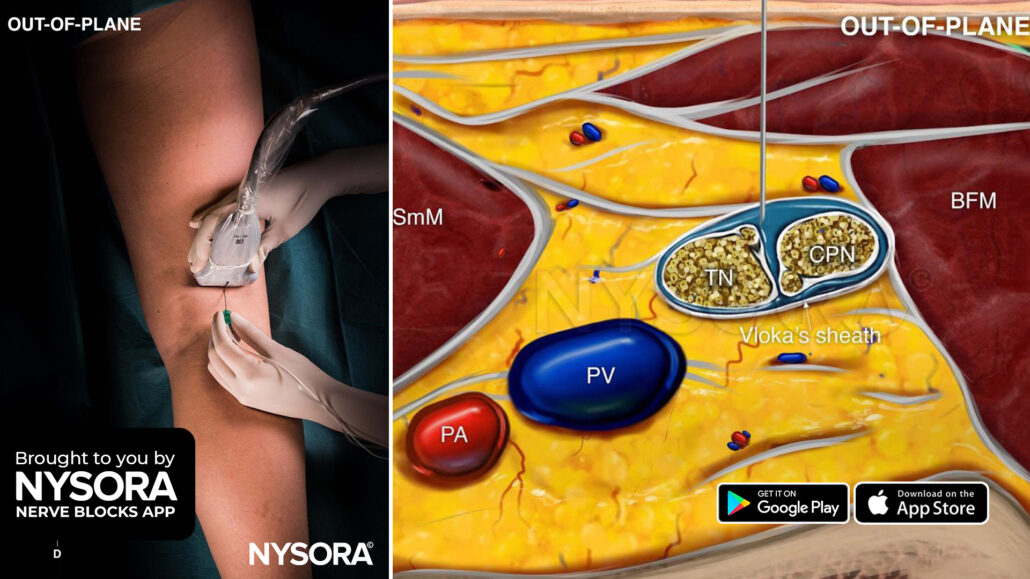
- Place the transducer transversely 3-5 cm above the popliteal fossa between the biceps femoris, and semimembranosus and semitendinosus tendons.
- Identify the sciatic nerve on ultrasound where the tibial (TN) and common peroneal (CPN) nerves start separating but are still within Vloka’s sheath.
- Insert the needle in-plane or out-of-plane into Vloka’s sheath between the TN and CPN, and inject 10-20 mL of local anesthetic.
Watch the video below to get a better picture of the process and see how the NYSORA Nerve Blocks App brings these instructions to life:
For more tips like these and the complete guide to the 60 most frequently used nerve blocks, download the Nerve Blocks App HERE. Don’t miss the chance to get the bestselling NYSORA Nerve Blocks App also in book format – the perfect study companion with the Nerve Blocks app!


