The Best Way to Learn or Teach: Infraclavicular Block
Let’s face it: Ultrasound-guided Regional Anesthesia practice, learning and teaching rely on visual information and videos. But then, books, e-books, and most websites on this topic are typically static and limited in visuals. That is why NYSORA’s Team developed a range of cognitive aids, such as Reverse Ultrasound Anatomy™ (RUA) illustrations and animation as a method to help learn and memorize relevant son-anatomy patterns needed for various ultrasound-guided procedures. To create a Reverse Ultrasound Anatomy™ animation, a team of sonographers, medical experts, illustrators, and animators work together to create simply the most powerful tools for learning ultrasound-guided procedures. In this newsletter, we will demonstrate how RUA™ is used to teach Infraclavicular Brachial Plexus Block, which is used for anesthesia and analgesia for surgeries in the arm, elbow, forearm, and hand.
“Watching the RUA animation where you can switch between ultrasound image and animation, and back is the most powerful tool to-date to learn ultrasonographic patterns. This quick exercise helps the user create ultrasonographic patterns that can be quickly recalled in clinical practice to facilitate the identification of the anatomical structures of importance to ultrasound-guided intervention. As such, it is extremely educational, vivid, and fun for teaching,” says Dr. Hadzic.
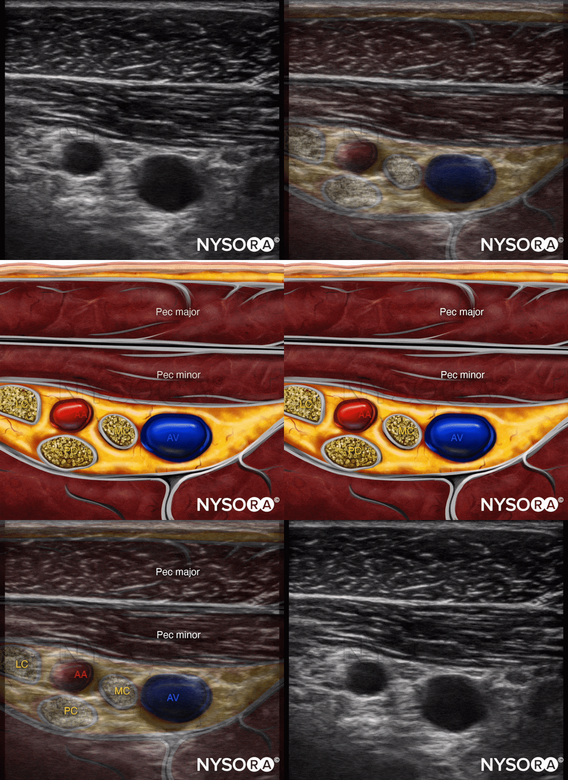
Here’s an example and an experiment for you. If you take a closer look at the Infraclavicular Brachial Plexus block Reverse Ultrasound Anatomy™ animation, you will notice that even with an excellent US image quality like in this video, it is difficult to recognize the individual cords of the brachial plexus. However, if you play the RUA™ a couple of times – back and forth, you will notice how now you immediately start recognizing all 3 cords even on the unlabeled ultrasound image. And that is exactly what the RUA™ project aims at – ultrasound pattern recognition to facilitate learning.

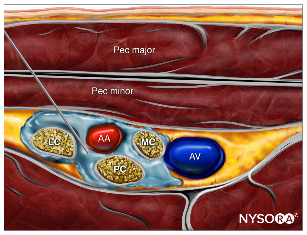
The next RUA™ illustration explains the goal of this block – the spread of local anesthetic around the axillary artery to the median, lateral, and posterior cords of the brachial plexus. A series of animations then animate the interaction of the needle with the tissue fasciae of the pectoralis muscles as it approaches the target of injection, behind the axillary artery.
The NYSORA Compendium of Regional Anesthesia is filled with material just like this, as it is a fun, comprehensive, and practical course built for visual learning. Get in-depth lessons, illustrations, animations, and clinical videos on local anesthesia, nerve blocks, intravenous, and spinal anesthesia. Immerse yourself in the expertly crafted learning aids and tips, take notes and make your own scripts with the content from the Compendium or elsewhere using the built-in note-taking tool. Your subscription will also give you access to the NYSORA LMS Activity Feed where you can read or create your own posts and tips on everything regional anesthesia while interacting with a community of inspiring practitioners.
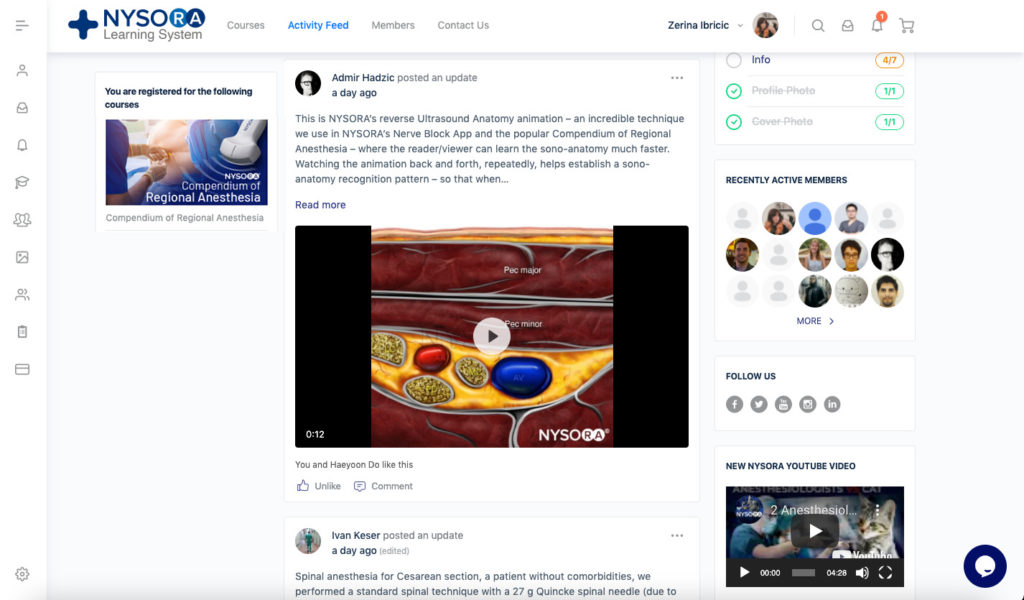
Watch this video to see how the NYSORA Compendium of Regional Anesthesia can revolutionize teaching and learning in your practice:
We created NYSORA’s Compendium to bring on a new wave in medical e-learning. Our excitement for creating this all-encompassing guide burned so brightly that an army of medical professionals, illustrators, animators, and video editors banded together to make the extensive course material reality over the course of years.
As a result, the Compendium of Regional Anesthesia is comparatively the most authoritative guide on regional anesthesia, from A to Z. You might be thinking “Oh, an ebook” – but that’s pretty far off. It may feel like an ebook on steroids – but it goes way beyond that. The compendium features literally thousands of visual aids and regular new additions and updates. But what we really paid attention to is that the Compendium is made for the visual learner.
