Ultrasound for Aortic Regurgitation
Point-of-Care Ultrasound (POCUS) is excellent for cardiac assessment. Major valvular diseases of the tricuspid, mitral, and aortic valves are also easy to identify.
Aortic regurgitation, also known by aortic insufficiency, is a significant valvular disease characterized by the backflow of blood from the aorta. This condition occurs when the aortic valve, which separates the left ventricle from the aorta, does not close properly. As a result, the heart needs to work harder to pump the volume overload.
Aortic regurgitation can have various underlying causes, with degenerative valve conditions, congenital defects, or other heart-related issues being the most common culprits. Upon diagnosis, it is essential to prioritize a formal transthoracic echocardiography, preferably from a certified healthcare professional.
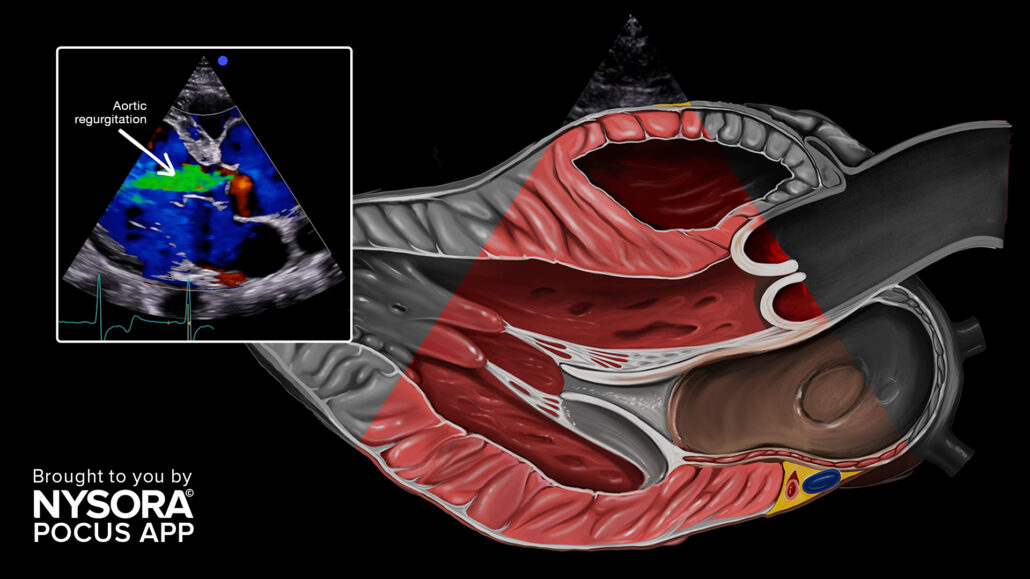
When evaluating aortic regurgitation through ultrasound, distinctive pathology characteristics to observe include:
- Aliasing with large jets, seen as multiple color blood flow on color Doppler.
- The regurgitation is severe if the jet occupies the majority (65%) of the left ventricle outflow tract.
- Jets that are not seen in the center of the valve tend to be underestimated and have to be considered severe until proven otherwise.
Aortic regurgitation.
Unleash the potential of POCUS with NYSORA’s POCUS App and elevate your practice, expand your capabilities, and deliver exceptional patient care. Download HERE.


