 Nerve Blocks App
 Anesthesia Assistant App
 Ultrasound Pain App
 Pain Medicine Assistant App
 POCUS App
 IV Access App
 MSK Knee App
 VetRA App
 Nerve Block Manual
 Regional Anesthesia Updates
 Anesthesiology Manual
 Anesthesiology Review
 Anesthesia Updates 2025
 Anesthesia Updates 2026
 Pediatric Anesthesia Updates
 Airway Management Updates
 US Interventional Pain Manual
 Pain Medicine Updates
 Mastering Difficult IV Access
 PACU Nursing Manual
 RA Veterinary Manual
 Books
Books About
About
This chapter will probably be the shortest. It was necessary to understand quietly, step-by-step, the elementary sign, the developed sign of interstitial syndrome, and the phenomenon of lung sliding. Lung ultrasound is a dynamic tool investigating a dynamic organ, using a sign, lung sliding, with no equivalent using traditional tools (X-rays and CT mainly). This allows to define two opposing kinds of interstitial syndrome, the transudative and the exudative interstitial syndrome.
In the BLUE-protocol, they were given short labels:
- The detection in a blue patient, at the anterior chest wall, of lung rockets associated with lung sliding (each word is important) has been called the B-profile
- The B’-profile is a B-profile with abolished lung sliding
This distinction follows the logic of the BLUE-protocol, by determining profiles using associations of signs [1]. When we write that lung sliding is “abolished” in the B’-profile, it can be completely, or quite completely abolished, with a 1-mm amplitude roughly. This distinction is of no relevance: both cases indicate a very impaired compliance. The question of pneumothorax makes no sense in the presence of any B-line. Note interestingly that videos are not mandatory if good quality M-mode data are provided.
The B-profile and mostly the B’-profile are seen independently from the type of ventilation (spontaneous or mechanical).
1. THE ULTRASOUND TRANSUDATIVE INTERSTITIAL SYNDROME (B-PROFILE)
Briefly, hemodynamic pulmonary edema generates transudate, a kind of oil preventing the lung to burn during an all-life breathing. Usually, the B-profile will be seen in hemodynamic pulmonary edema.
2. THE ULTRASOUND EXUDATIVE INTERSTITIAL SYNDROME (B’-PROFILE)
Inflammatory diseases generate exudate, fibrin, and various viscous stuffs, which may stick the lung to the wall. The B’-profile will prove a highly specific sign of pneumonia/ARDS. (Fig. 1)
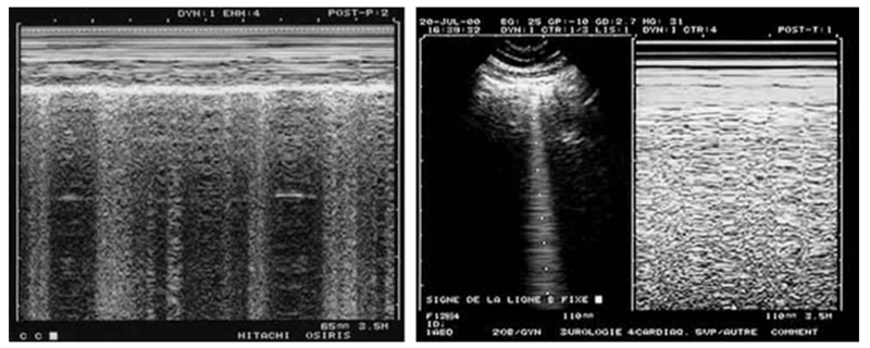
Fig. 1 This figure is devoted to show that static images can indicate dynamic phenomena. To the left, the alternation of vertical hyperechoic lines shows that B-lines come and go through the shooting line of the M-mode: lung sliding is present. Note also the marked dyspnea in the Keye’s space: patient likely not intubated. To the right, the M-mode shows a homogeneous field below the Keye’s space, indicating that the B-line taken by the shooting line is standstill.
3. THE LANGUAGE OF THE BLUE-PROTOCOL, ITS MAIN PRINCIPLE
The label “B-profile” should be understood, like in aviation language, where speed is life, and where most pieces of information must be said in a minimal time. Instead of “B-profile,” one could have said: “the pattern with ten features, which is a comet-tail artifact, arising from the pleural line, moving with lung sliding, usually long, usually well-defined, usually erasing A-lines, usually hyperechoic, multiple (three or more) in one longitudinal scan, disseminated to the anterior bilateral chest wall, and moving with a preserved lung sliding,” a little long for being adopted. Words are the principle of any language. In usual conditions, the “B-profile” and the “B’-profile” are recognized in a few seconds or less. One of the immediate therapeutic outcomes is the suggestion of CPAP in the case of a B-profile and direct intubation in the case of a B’-profile, as seen in the detailed chapters.