 Nerve Blocks App
 Anesthesia Assistant App
 Ultrasound Pain App
 Pain Medicine Assistant App
 POCUS App
 IV Access App
 MSK Knee App
 VetRA App
 Nerve Block Manual
 Regional Anesthesia Updates
 Anesthesiology Manual
 Anesthesiology Review
 Anesthesia Updates 2025
 Anesthesia Updates 2026
 Pediatric Anesthesia Updates
 Airway Management Updates
 US Interventional Pain Manual
 Pain Medicine Updates
 Mastering Difficult IV Access
 PACU Nursing Manual
 RA Veterinary Manual
 Books
Books About
About
 Back to News
 9 min read

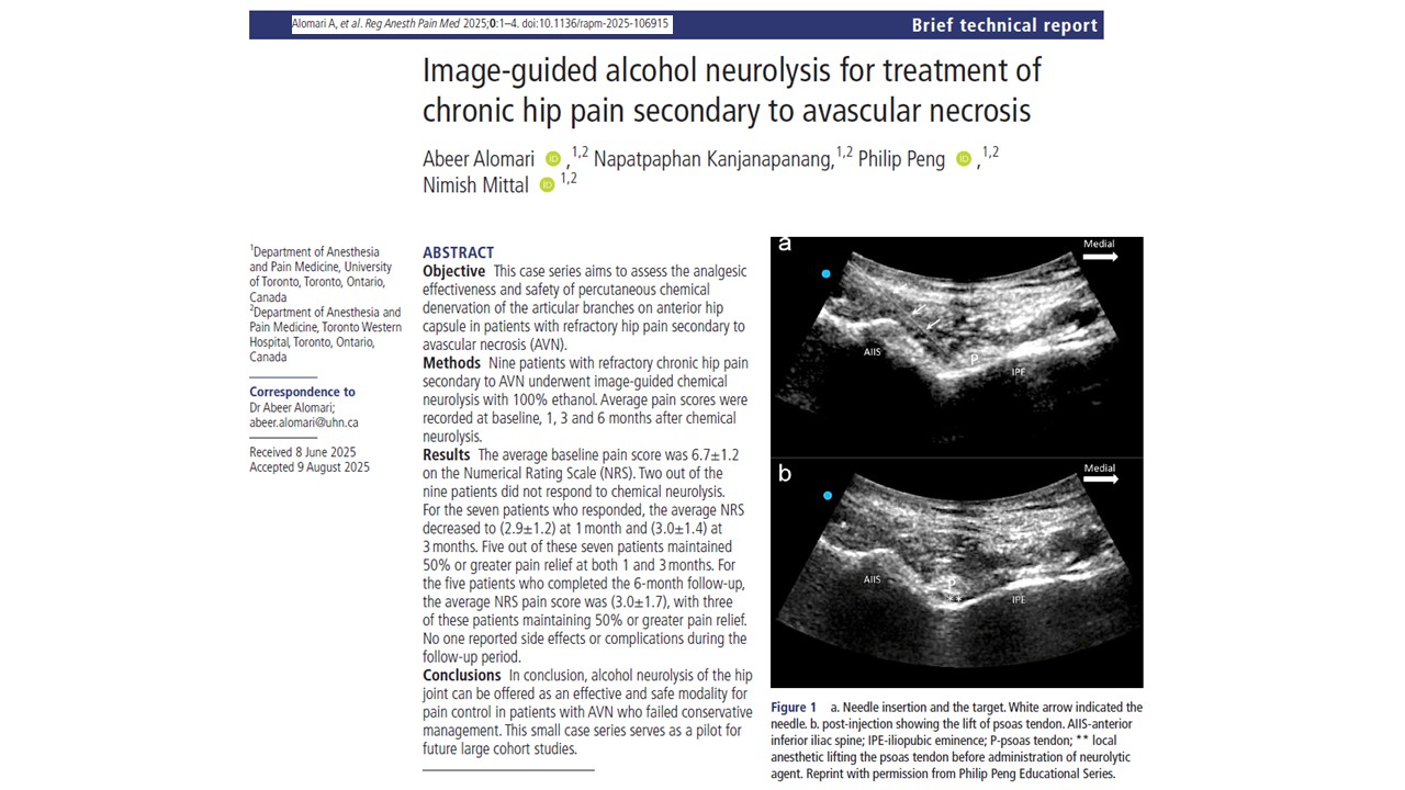
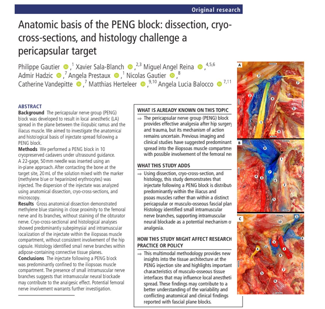
A new anatomical and histological study is challenging a central assumption about how the pericapsular nerve group (PENG) block works.
Published in Regional Anesthesia & Pain Medicine in 2026, the study by Gautier and colleagues found that injectate administered during a PENG block was located predominantly within the iliacus and iliopsoas muscle compartments rather than consistently spreading along a distinct pericapsular plane toward the anterior hip capsule.
The findings could help explain two observations that have puzzled clinicians: why the PENG block can provide effective analgesia even when expected pericapsular spread is not demonstrated, and why quadriceps weakness can sometimes occur despite the technique being developed as a motor-sparing regional anesthetic block.
Importantly, the study does not demonstrate that the PENG block is ineffective. Instead, it raises questions about its anatomical mechanism of action.
Why the anatomy of the PENG block matters
The PENG block was introduced as a regional anesthesia technique for hip pain, particularly in patients undergoing hip surgery or experiencing hip fracture.
The original concept was to target articular branches supplying the anterior hip capsule, including contributions from the:
- Femoral nerve
- Obturator nerve
- Accessory obturator nerve
Local anesthetic was expected to spread through a plane between the iliopubic ramus and the overlying iliacus muscle, potentially reaching these articular branches while limiting blockade of the main femoral nerve.
That theoretical selectivity helped establish the PENG block’s reputation as a potentially motor-sparing hip block.
However, clinical reports of quadriceps weakness following PENG blocks and imaging studies showing injectate within the iliopsoas musculature have raised questions about this model.
The new investigation examines those questions at the macroscopic, cross-sectional and microscopic levels.
How the researchers studied PENG block spread
The investigators examined 15 hips from 10 cryopreserved cadaveric specimens.
Rather than relying on a single method of evaluating injectate distribution, the researchers combined three complementary approaches:
- Gross anatomical dissection in three hips
- Cryo-cross-sectional analysis in eight hips
- Histological analysis in four hips, including two uninjected controls
Ultrasound-guided PENG blocks were performed according to the originally described technique.
A 22-gauge, 50 mm needle was advanced in-plane from lateral to medial. After bone contact at the target site, 20 mL of injectate was administered.
Methylene blue was used for macroscopic and cryo-cross-sectional evaluation. For part of the histological investigation, heparinized erythrocytes were used as a particulate marker capable of showing where the bulk injectate was deposited microscopically.
This multimodal methodology was important because each technique answers a slightly different anatomical question.
Where did the injectate actually go?
The principal finding was striking: injectate was predominantly associated with the iliacus and iliopsoas muscle compartments.
Cryo-cross-sections showed methylene blue along the deep aspect of the iliacus and extending into the muscle.
At the level of the psoas tendon near the pelvic brim, staining remained enclosed within the iliopsoas compartment.
At the level of the femoral head and anterior hip capsule, the investigators again observed staining closely associated with the iliopsoas musculature, without consistent extension into the hip capsule.
In other words, the results did not support preferential distribution through a clearly defined musculo-osseous plane extending toward the anterior hip capsule.
This challenges the traditional description of the PENG block as primarily a pericapsular injection.
Histology provides a closer look
The histological component of the study provided particularly important information about the microanatomy of the injection site.
The investigators identified a capsule-periosteum complex composed of dense collagenous connective tissue.
Within the iliacus muscle, muscle fibers were organized into fascicles surrounded by connective tissue structures, including the perimysium and epimysium. Adipose-containing connective tissue spaces also contained blood vessels and small nerve branches.
Following injection, the erythrocyte marker was found predominantly:
- Within the epimysial compartment
- Along connective tissue planes between perimysial septa
- Within intramuscular adipose-containing spaces
- Near small blood vessels and nerve branches
The marker was not identified within the dense collagen fibers of the capsule-periosteum complex.
This observation suggests that injected fluid preferentially follows lower-resistance, adipose-containing connective tissue pathways rather than freely crossing dense collagenous structures.
Could the PENG block work through intramuscular nerves?
One of the most intriguing findings was the identification of small nerve branches within the intramuscular connective tissue spaces containing the injectate marker.
This raises an alternative or complementary explanation for PENG analgesia.
Rather than depending exclusively on local anesthetic reaching articular nerves immediately adjacent to the hip capsule, the block may partly act through neural structures encountered within the iliopsoas muscle compartment.
The authors therefore propose intramuscular neural blockade as a potential contributor to analgesia.
This hypothesis could help reconcile the apparent contradiction between effective clinical analgesia and anatomical studies that do not consistently demonstrate the expected pericapsular spread.
It remains a proposed mechanism, however, rather than definitive proof of which nerves are responsible for the clinical effect.
What happened around the femoral nerve?
The relationship between the injectate and the femoral nerve may have important clinical implications.
Gross anatomical dissection showed methylene blue in close proximity to the femoral nerve and several of its branches along the anterior surface of the iliopsoas muscle.
Cryo-cross-sectional analysis also demonstrated injectate near the femoral nerve region.
In contrast, dye was not observed around the obturator nerve within the obturator canal.
This distribution raises the possibility that local anesthetic deposited during a PENG block can diffuse toward the femoral nerve.
The authors emphasize an important distinction: visible dye or particulate markers show bulk injectate distribution, but individual local anesthetic molecules may diffuse beyond the boundaries demonstrated by those markers.
Therefore, the absence of visible marker around a nerve does not necessarily prove that pharmacologically active local anesthetic cannot reach it.
Could this explain quadriceps weakness?
Potential femoral nerve involvement is particularly relevant because the femoral nerve supplies motor innervation to the quadriceps.
Quadriceps weakness has previously been reported after PENG blocks, creating a potential problem for a technique often described as motor sparing.
The new anatomical findings provide a plausible explanation.
If local anesthetic spreads or diffuses from the iliopsoas compartment toward the femoral nerve or its motor branches, quadriceps function could potentially be affected.
This does not mean every PENG block will produce femoral nerve blockade or clinically significant motor weakness.
It does mean that motor sparing should not automatically be assumed solely from the intended anatomical target.
Further clinical research is needed to establish how frequently this occurs and how factors such as local anesthetic volume, concentration, injection location and individual anatomy influence the risk.
What did the study find about the obturator nerve?
The original PENG concept includes blockade of articular branches associated with the obturator nerve.
In this cadaveric investigation, however, gross anatomical dissection did not demonstrate methylene blue around the obturator nerve within the obturator canal.
This does not establish that obturator-derived articular innervation can never be affected by a PENG block.
The study did not specifically trace every individual articular branch from the femoral, obturator and accessory obturator nerves to determine whether local anesthetic reached them.
The findings instead show that bulk injectate did not follow a simple pathway from the injection site to the main obturator nerve.
Why different anatomical studies can produce different answers
The study also highlights an important methodological issue in regional anesthesia research.
A tissue that appears stained during gross dissection does not necessarily represent the primary compartment occupied by the injected solution.
Methylene blue can diffuse beyond the area containing the bulk of the injectate. This could create superficial staining that appears to indicate spread into a particular anatomical region.
Cryo-cross-sectioning preserves anatomical relationships differently, while histological markers such as erythrocytes can provide a more precise indication of where bulk fluid has traveled at the microscopic level.
The researchers therefore caution against interpreting visible periosteal staining alone as proof that a substantial volume of injectate has entered a pericapsular or musculo-osseous plane.
Key findings for clinicians
The study’s most clinically relevant observations include:
- Injectate was predominantly distributed within the iliacus and iliopsoas muscle compartments
- Consistent spread to the anterior hip capsule was not demonstrated
- Histology showed injectate in subepimysial and intramuscular connective tissue pathways
- Small nerve branches were present close to the intramuscular injectate
- Gross dissection demonstrated staining close to the femoral nerve and several of its branches
- The obturator nerve was not stained within the obturator canal
- Diffusion toward the femoral nerve could potentially contribute to quadriceps weakness
- The anatomical mechanism responsible for PENG analgesia remains incompletely understood
A changing anatomical model of the PENG block
The most important message from the study may be conceptual.
A successful regional anesthetic block does not necessarily work exactly as its original anatomical model predicts.
Gautier and colleagues found that a conventional PENG injection predominantly entered the iliacus and iliopsoas muscle compartments rather than consistently reaching a distinct pericapsular plane. Histology further revealed small neural structures within the same connective tissue pathways occupied by the injectate.
At the same time, the close relationship between the injectate and femoral nerve raises a clinically important question about motor blockade.
The findings therefore shift attention away from a purely pericapsular model and toward the microanatomy of the iliopsoas compartment.
Further in vivo studies will be necessary to determine how injection volume and anatomical distribution relate to analgesic efficacy and quadriceps function.
Reference: Gautier P et al. Anatomic basis of the PENG block: dissection, cryo-cross-sections, and histology challenge a pericapsular target. Reg Anesth Pain Med. Published online July 24, 2026.
Download the Nerve Blocks App HERE for instant access to step-by-step guidance on the PENG block and other regional anesthesia techniques. Prefer a physical copy? The bestselling NYSORA Nerve Blocks App is available in book format – an essential resource for mastering nerve blocks! And for a digital learning experience, check out the Nerve Blocks Module on NYSORA360!