 Nerve Blocks App
 Anesthesia Assistant App
 Ultrasound Pain App
 Pain Medicine Assistant App
 POCUS App
 IV Access App
 MSK Knee App
 VetRA App
 Nerve Block Manual
 Regional Anesthesia Updates
 Anesthesiology Manual
 Anesthesiology Review
 Anesthesia Updates 2025
 Anesthesia Updates 2026
 Pediatric Anesthesia Updates
 Airway Management Updates
 US Interventional Pain Manual
 Pain Medicine Updates
 Mastering Difficult IV Access
 PACU Nursing Manual
 RA Veterinary Manual
 Books
Books About
About
 Back to News
 7 min read
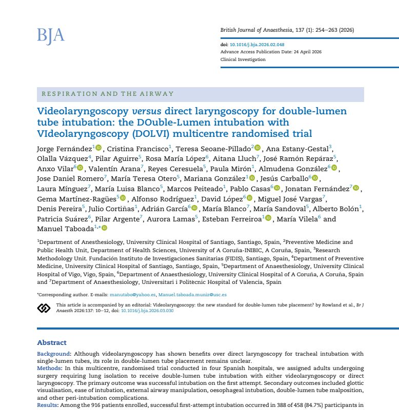
A large multicentre randomised trial has found that videolaryngoscopy improves first-attempt success during double-lumen tube intubation compared with conventional direct laryngoscopy in adults undergoing surgery requiring lung isolation.
The DOuble-Lumen intubation with VIdeolaryngoscopy (DOLVI) trial enrolled 916 patients across four hospitals in Spain. First-attempt intubation was successful in 84.7% of patients using videolaryngoscopy compared with 76.9% using direct laryngoscopy, an absolute difference of 7.9 percentage points.
Published in the British Journal of Anaesthesia in 2026, the findings provide evidence that videolaryngoscopy with a standard Macintosh-type blade can improve several clinically important aspects of double-lumen tube placement without prolonging intubation time.
Why double-lumen tube intubation is challenging
Double-lumen tubes are widely used when lung isolation is required during thoracic surgery. They allow independent ventilation of the lungs and facilitate procedures in which one lung needs to be collapsed to improve surgical exposure.
However, inserting a double-lumen tube is more technically demanding than conventional single-lumen tracheal intubation.
Several characteristics contribute to this difficulty:
- Larger diameter: double-lumen tubes are bulkier than conventional tracheal tubes.
- Greater rigidity: their construction makes manipulation more difficult.
- Fixed bronchial curvature: the bronchial component must be correctly orientated during placement.
- Additional rotation: after the bronchial cuff passes through the vocal cords, the tube typically requires a 90-degree rotation before advancement.
- Precise positioning: the bronchial lumen must ultimately align with the appropriate main bronchus.
Failure on the first attempt can result in repeated laryngoscopy and potentially increase the risks of prolonged apnoea, hypoxemia, hemodynamic instability, airway trauma and tube malposition.
Videolaryngoscopy provides an indirect camera view of the glottis and has demonstrated advantages for single-lumen tracheal intubation. Whether those advantages translate to double-lumen tubes has been less certain.
How the DOLVI trial was conducted
The prospective, multicentre, unblinded, parallel-group randomised trial was conducted between July 2024 and October 2025.
Adults aged 18 years or older undergoing elective or urgent surgery requiring double-lumen tube intubation were eligible.
A total of 995 patients were assessed for eligibility and 916 underwent randomisation. Patients were allocated equally between the two strategies:
- 458 patients: videolaryngoscopy
- 458 patients: direct laryngoscopy
The median patient age was 67 years and approximately 61% were men.
Most procedures were elective. Lobectomy or segmentectomy represented the largest surgical category, followed by wedge resection.
Standard Macintosh-type blades were used
An important feature of the trial was the type of videolaryngoscope studied.
Patients assigned to videolaryngoscopy were intubated primarily using videolaryngoscopes equipped with standard Macintosh-type geometry blades, rather than hyperangulated blades.
Of the 458 patients allocated to videolaryngoscopy, 457 underwent the first attempt with a standard-geometry blade. The devices included McGrath MAC and Storz C-MAC videolaryngoscopes.
Only one patient received a hyperangulated blade.
This distinction is clinically important because the trial findings cannot automatically be extrapolated to hyperangulated videolaryngoscopes.
First-attempt success increased with videolaryngoscopy
The primary outcome was successful tracheal intubation on the first attempt.
Success required placement of the double-lumen tube in the trachea following a single insertion of the laryngoscope blade and a single advancement of the tube through the vocal cords, with confirmation by waveform capnography.
The results showed:
- Videolaryngoscopy: 388/458 patients, or 84.7%
- Direct laryngoscopy: 352/458 patients, or 76.9%
- Absolute risk difference: 7.9 percentage points
- 95% confidence interval: 2.8–12.8 percentage points
- P-value: 0.003
The findings therefore demonstrated a statistically significant improvement in first-attempt success with videolaryngoscopy.
The benefit was generally consistent across prespecified subgroups, hospitals, operator experience and patient characteristics.
Glottic visualisation was substantially better
Videolaryngoscopy also produced a clear improvement in visualisation of the glottis.
Good glottic visualisation, defined as modified Cormack–Lehane grade I or IIa, was achieved in:
- 93.7% with videolaryngoscopy
- 80.8% with direct laryngoscopy
This represented an absolute difference of 12.9 percentage points.
Grade I view was obtained in 79.3% of patients undergoing videolaryngoscopy compared with 59.2% undergoing direct laryngoscopy.
Better visualisation was accompanied by less loss of the glottic view as the double-lumen tube was advanced.
Intubation was easier for operators
“Easy intubation” was defined by the investigators as successful first-attempt intubation combined with favourable glottic visualisation.
This occurred in:
- 81.9% of videolaryngoscopy cases
- 68.6% of direct laryngoscopy cases
The absolute difference was 13.3 percentage points.
Operators also reported fewer moderately or severely difficult intubations with videolaryngoscopy.
External airway manipulation was required in 28.6% of patients receiving videolaryngoscopy compared with 40.6% receiving direct laryngoscopy.
The need to switch the intubating device was also markedly lower: 0.9% versus 9.2%, respectively.
Importantly, these improvements did not come at the cost of a longer procedure. Median intubation duration was approximately 22 seconds with videolaryngoscopy and 23 seconds with direct laryngoscopy.
Fewer oesophageal intubations and tube malpositions
The trial also examined several exploratory safety outcomes.
Oesophageal intubation occurred in:
- 1.7% with videolaryngoscopy
- 4.6% with direct laryngoscopy
Double-lumen tube malposition occurred in:
- 7.6% with videolaryngoscopy
- 12.4% with direct laryngoscopy
The differences for both outcomes were statistically significant.
In contrast, rates of airway injury, hypoxemia, hypotension, postoperative sore throat and hoarseness were similar between groups. The trial was not powered to detect differences in rare adverse events.
Why blade geometry may matter
The reduction in double-lumen tube malposition is particularly noteworthy because previous studies have produced conflicting results.
The DOLVI investigators suggest that blade geometry may partly explain the difference.
Hyperangulated videolaryngoscopes can provide excellent views of the glottis, but their pronounced curvature can make advancement of a large and relatively rigid double-lumen tube more difficult.
The bronchial tip may encounter the anterior tracheal wall or arytenoid structures, potentially complicating advancement and rotation.
In DOLVI, standard Macintosh-type videolaryngoscope blades provided a gentler curvature and a more linear trajectory towards the glottis.
The investigators propose that this configuration may combine improved visualisation with easier double-lumen tube advancement, potentially contributing to the lower malposition rate observed in the trial.
What could the results mean for clinical practice?
The findings support consideration of standard-geometry videolaryngoscopy as an initial approach for double-lumen tube placement in patients requiring lung isolation.
The potential clinical advantages demonstrated by the trial include:
- Higher first-attempt success
- Better glottic visualisation
- Fewer repeat intubation attempts
- Less external airway manipulation
- Less frequent loss of the glottic view
- Fewer oesophageal intubations
- Lower double-lumen tube malposition
- Less frequent need to change the intubating device
- No meaningful increase in intubation time
These findings are especially relevant because approximately two-thirds of the trial’s intubations were performed by anesthesia residents, although the participating centres had substantial experience with both videolaryngoscopy and direct laryngoscopy.
How is a double-lumen tube placed?
In simplified terms, the technique used in the trial involved several stages:
- Visualise the glottis: the vocal cords are identified using either the assigned videolaryngoscope or direct laryngoscope.
- Advance the tube: the pre-shaped double-lumen tube is passed until its bronchial cuff has crossed the vocal cords.
- Remove the stylet: the malleable stylet is removed after the bronchial cuff passes the cords.
- Rotate the tube: left-sided tubes are rotated approximately 90 degrees anticlockwise, whereas right-sided tubes are rotated approximately 90 degrees clockwise.
- Advance into position: the tube is gently advanced until mild resistance is encountered.
- Inflate the cuffs and ventilate: the tracheal cuff is inflated followed by the bronchial cuff, and ventilation is initiated.
The study’s primary endpoint focused specifically on successful passage into the trachea. Further manipulation or rotation required to achieve bronchial positioning after the tube passed the vocal cords did not constitute another intubation attempt.
The bottom line
The DOLVI trial provides large, multicentre randomised evidence supporting videolaryngoscopy for double-lumen tube placement during surgery requiring lung isolation.
Compared with conventional Macintosh direct laryngoscopy, videolaryngoscopy with a standard Macintosh-type blade increased first-attempt success, substantially improved glottic visualisation and reduced several measures of procedural difficulty.
It was also associated with fewer oesophageal intubations and double-lumen tube malpositions, without increasing intubation time or the overall incidence of several other peri-intubation complications.
The investigators conclude that their findings suggest videolaryngoscopy is the preferable initial approach for airway management in patients requiring lung isolation.
Reference: Fernández J et al. Videolaryngoscopy versus direct laryngoscopy for double-lumen tube intubation: the DOuble-Lumen intubation with VIdeolaryngoscopy (DOLVI) multicentre randomised trial. Br J Anaesth. 2026;137:254-263.
Read more about recent evidence like this in NYSORA’s Anesthesia Assistant app. Simulate case, check dosages and search through NYSORA knowledge database!